
Abstract
Autophagy is an innate immune defense against bacterial invasion. Recent studies show that two adaptor proteins, p62 and NDP52, are required for autophagy of the bacterial pathogen Salmonella enterica serovar Typhimurium (S. Typhimurium). However, it is not known why two different adaptors are required to target the same bacterial cargo to autophagy. Here we show that both adaptors are recruited to bacteria with similar kinetics, that they are recruited to bacteria independently of each other, and that depletion of either adaptor leads to impairment of antibacterial autophagy. Depletion of both adaptors does not synergistically impair autophagy, indicating they act in the same pathway. Remarkably, we observed that these adaptors do not colocalize, but rather form non-overlapping microdomains surrounding bacteria. We conclude that p62 and NDP52 act cooperatively to drive efficient antibacterial autophagy by targeting the protein complexes they coordinate to distinct microdomains associated with bacteria.
Acknowledgements
John H. Brumell, Ph.D., is the recipient of an Investigators in Pathogenesis of Infectious Disease Award from the Burroughs Wellcome Fund. We thank Dr. John Kendrick-Jones (University of Cambridge, MRC) for kindly providing the NDP52-GFP construct, and to members of the Brumell laboratory for technical assistance and critical reading of this manuscript.
Figures and Tables
Figure 1 The ubiquitin-binding adaptors, p62 and NDP52, are recruited to S. typhimurium with the same kinetics. (A) HeLa cells were infected with S. typhimurium expressing RFP. Cells were fixed at the indicated time and stained with antibody to p62 or NDP52. The percentage of p62+ or NDP52+ bacteria was enumerated by fluorescence microscopy. At least 100 bacteria were counted for each time point. The experiment was conducted two times and error bars represent the range. (B) HeLa cells were infected for 1 h, fixed and stained for p62 and NDP52. The percentage of p62 colocalizing with NDP52+ bacteria (left part) and the percentage of NDP52 colocalizing with p62+bacteria (right part) were enumerated by fluorescence microscopy. At least 100 bacteria were counted for each condition. The average ± SD for three independent experiments is shown. (C) HeLa cells were transfected with p62-mCherry and ND P52-GFP or p62-mCherry and GFP. Then cells were infected with S. typhimurium (1 h), or treated with rapamycin (2 h), as indicated. HeLa lysates were immunoprecipitated (IP) with GFP antibody. Lysates and precipitates were then blotted (WB) for p62 and GFP. The lysate blot also contains the GFP alone band (27 kDa) which is not depicted in the figure. (D) HeLa cells were coimmunostained with p62 and NDP52 antibodies. Insets show higher magnification in the area indicated with an arrow.
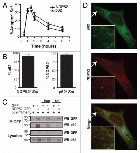
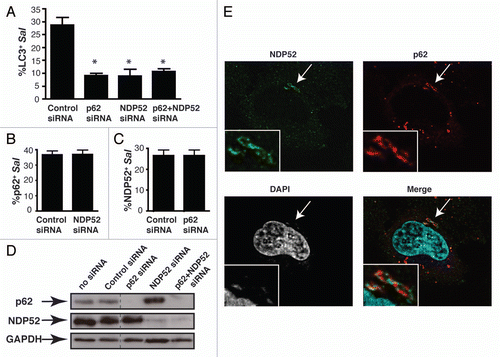
Figure 2 p62 and NDP52 are recruited independently to bacteria-associated microdomains to promote autophagy of S. typhimurium. (A) HeLa cells were treated with control, p62, NDP52 or p62 + NDP52 siRNA for 48 h and also transfected with GFP-LC3 for 24 h. Cells were then infected with S. typhimurium and fixed at 1 h p.i. Cells were then stained for external and internal bacteria. The percentage of LC3+ intracellular bacteria was enumerated by fluorescence microscopy. At least 100 bacteria were counted for each condition. The average ± SD is shown for three independent experiments. Asterisk denotes p value <0.001 calculated by two-tailed Student's t-test. (B and C) HeLa cells were transfected with the indicated siRN A and then infected with S. typhimurium for 1 h. The percentage of p62+ bacteria (B) and the percentage of NDP52+ bacteria (C) were enumerated by fluorescence microscopy. At least 100 bacteria were counted for each condition. The average ± SD for four independent experiments is shown. (D) Protein lysates from siRNA-treated cells were harvested and analyzed by western blot using antibodies to p62 and NDP52. GAPDH was used as a loading control. (E) HeLa cells were infected with S. typhimurium and fixed at 1 h p.i. Cells were then co-immunostained with p62 and NDP52 antibodies and stained with DAPI to label DNA. Insets show higher magnification in the boxed areas.
Addendum to: