
Abstract
Purpose: To investigate the frequency of xenotropic murine leukemia virus (MLV) presence in human cell lines established from mouse xenografts and to search for the evidence of horizontal viral spread to other cell lines. Methodology: We examined xenograft tumor cell lines from 7 independent laboratories and 128 non-xenografted tumor cell lines. Cell line DNA was examined for mouse DNA contamination, and by three Taqman qPCR assays targeting the gag, env or pol regions of MLV. Sequencing was used for viral strain identification. Supernatant fluids were tested for reverse transcriptase (RT) activity. Results: Six of 23 (26%) mouse DNA free xenograft cultures were strongly positive for MLV and their sequences had greater than 99% homology to known MLV strains. Four of five available supernatant fluids from these viral positive cultures were strongly positive for RT activity. Three of these supernatant fluids were studied to confirm the infectivity of the released virions for other human culture cells. Of the 78 non-xenograft derived cell lines maintained in the xenograft culture-containing facilities, 13 (17%) were positive for MLV, including XMRV, a virus strain first identified in human tissues. By contrast, all 50 cultures maintained in a xenograft culture-free facility were negative for viral sequences. Conclusions: Human cultures derived after mouse xenografting frequently contain and release highly infectious xenotropic MLV viruses. Laboratories working with xenograft-derived human cultures should be aware of the risk of contamination with potentially biohazardous human-tropic mouse viruses and their horizontal spread to other cultures.
Disclosure of Potential Conflicts of Interest
No potential conflicts of interest were disclosed.
Acknowledgments
We thank Misty Watson, Jason Toombs, Laura Sullivan, Juliet Rivera, Dr. Chun-Xian Huang, Dr. Puja Gupta, Dr. Yi Li, Dr. Philip E. Thorpe and Dr. Yi Yin at UT Southwestern Medical Center for their kind help in procurement of mouse tissues, xenografts and xenograft cell lines, Sara Murphy at Johns Hopkins University School of Medicine for kindly preparing the DNA of cell lines from Dr. Charles M. Rudin Laboratory, Dr. Charles Sawyers and John Wongvipat at Memorial Sloan-Kettering Cancer Center for generously providing LAPC-4 cell line and its related discussions. We acknowledge the assistance from Dr. James Eshleman at Johns Hopkins University School of Medicine in establishing the pancreatic carcinoma cell line.
Funding was provided by the Texas Specialized Program of Research Excellence in Lung Cancer (P50CA70907) and the Early Detection Research Network (U01CA084971), National Cancer Institute and the Canary Foundation (Palo Alto, CA).
Figures and Tables
Figure 1 Detection of MLV-related sequences by multiple MLV specific Taqman qPCR assays. (A and B) Validation of MLV qGag and qGPP assays. XMRV VP62 containing plasmids as standard references were serially diluted in an range of 1 × 10⁷ copies to one copy/reaction (copies/rxn) with salmon sperm carrier genomic DNA (100 ng/rxn) and detected in a quantitative range of over 6 logs by MLV qGag or qGPP PCR. The lowest detection limit is one to five copies of MLV 100 ng DNA. The validation of MLV qEnv for xenotropic MLV detection was described previously (see Materials and Methods). Note: validation of the third MLV assay, MLV qEnv, has already been published. (C) Detection of MLV N417 provirus by two viral probes in the DNA of late passage of MLV N417 virus-containing NCI-H60 culture. The DNA of late passage of NCI-H60 (50 ng) culture was strongly positive by qGPP (green) and qEnv (red) probes but not qGag (blue). These findings are consistent with the specificity of MLV N417 viral sequence present in the reference NCI-N417 cell line (Fig. S1 and ). By contrast, the viral free early passage of NCI-H60 culture, pooled human WBC DNA and water controls were all negative by all three MLV qPCR assays. (D) Detection of MLV DG-75 provirus by all three viral probes in CAK1 pancreatic carcinoma xenograft line. The DNA of CAK1 cells (100 ng) was strongly positive by all three MLV qPCR assays as indicated as qGag (blue), qGPP (green) and qEnv (red) (). These findings are consistent with the specificity of the sequences which were identified in CAK1 and homologous (> 99.0%) with MLV DG-75 virus (Fig. S1 and 5). Water controls in triplets were negative in each assay.
Rxn = reaction.
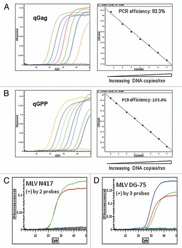
Figure 2 Identification of mouse DNA in human pancreatic carcinoma cultures established from mouse xenografts by PCR. A sensitive PCR assay specific for mouse GAP DH and its pseudogenes was used (Fig. S2 and Materials and Methods). The genomic DNA of human pancreatic carcinoma XCL1 and XCL3 xenograft cultures were found positive for mouse DNA. By contrast, the DNA of human pancreatic carcinoma CAK1 and human colorectal carcinoma RKO culture as well as the control pooled human WBC DNA were free mouse DNA (see Materials/Methods and Table S1–3).
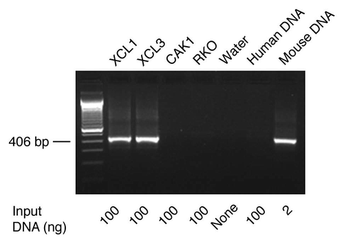
Figure 3 Infectivity of supernatant fluids of MLV positive xenograft derived cell lines. Five previously XMLV virus-free cultures (2 NSCLC lines: NCI-H1299 and NCI-H460; two SCLC lines: NCI-H69 and NCI-H1092, and an immortalized human bronchial epithelial cell culture HBEC3K-TR53 [HBEC3]), were infected with the supernatant fluids (250 µl) from 3 XMLV-positive xenograft derived cell lines (LAP C-4, NCI-N417 and 1065met) (), which had the reverse transcriptase (RT) activities of 5.7 × 10⁴ nU/µl, 3.1 × 10⁵ nU/µl and 1.8 × 10⁵ nU/µl, respectively. The supernatants collected at days 5 and 14 post-infection were examined for the RT activity (see Materials/Methods). The supernatants from the infected NSCLC cell line NCI-H460 showed minimal or no RT activities for all three viruses on day 5, 14 and 18 post-infection (data not shown).
![Figure 3 Infectivity of supernatant fluids of MLV positive xenograft derived cell lines. Five previously XMLV virus-free cultures (2 NSCLC lines: NCI-H1299 and NCI-H460; two SCLC lines: NCI-H69 and NCI-H1092, and an immortalized human bronchial epithelial cell culture HBEC3K-TR53 [HBEC3]), were infected with the supernatant fluids (250 µl) from 3 XMLV-positive xenograft derived cell lines (LAP C-4, NCI-N417 and 1065met) (Table 1), which had the reverse transcriptase (RT) activities of 5.7 × 10⁴ nU/µl, 3.1 × 10⁵ nU/µl and 1.8 × 10⁵ nU/µl, respectively. The supernatants collected at days 5 and 14 post-infection were examined for the RT activity (see Materials/Methods). The supernatants from the infected NSCLC cell line NCI-H460 showed minimal or no RT activities for all three viruses on day 5, 14 and 18 post-infection (data not shown).](/cms/asset/ba784465-8e4f-49e0-8348-01c42e1d9aa9/kcbt_a_10915955_f0003.gif)
Table 1 Identification of xenotropic murine leukemia viruses (XMLV) and MLV-related viruses in xenograft cell lines
Table 2 Detection of mouse leukemia virus (MLV) in both early and later passage of prostate LAP C-4 cell line
Table 3 Frequent detection of murine leukemia virus (MLV) contamination of non-xenograft human cultures
Table 4 Characterization of murine leukemia viruses (MLV) detected in human non-xenograft cultures in xenograft culture laboratoriesTable Footnote1