Abstract
Purpose: To investigate the possible role of microRNAs in the resistance to platinum based chemotherapy in non-small cell lung cancer (NSCLC), explore their potential role and find potential biomarkers for prediction of the response to platinum.
Patients and methods: Microarray was employed to compare the expression of miRNAs between A549 and A549/CDDP cells. The effect of a differently expressed miRNA (miR-21) was examined on the sensitivity of cells to platinum. MiR-21 expression in NSCLC tumor tissues and matched plasma sample was also analyzed by Real-time PCR.
Results: 21 miRNAs were deregulated in A549/CDDP. Increased miR-21 expression significantly increased the resistance of A549 cell to platinum, whereas reduced miR-21 decreased the resistance of A549/CDDP cell. This finding was further validated in the tissue samples of 58 patients and it was found that miR-21 expression was significantly increased in platinum based chemotherapy-resistant patients (n = 58, p = 0.000). And increased miR-21 expression was associated with the shorter DFS (p = 0.008). Among these 58 patients, 32 had the corresponding plasma samples and similar tendencies were detected in 68.75% patients. Finally, transfection of A549/CDDP with anti-miR-21 increased the expression of PTEN and decreased Bcl-2. In contrast, pre-miR-21 decreased the expression of PTEN and increased Bcl-2 in A549.
Conclusion: Our data suggests that the expression level of miR-21 in tumor tissue and plasma might be used as a biomarker to predict adjuvant platinum based chemotherapy response and disease free survival in patients with NSCLC. Thus, it may serve as a novel therapeutic target to modulate platinum-based chemotherapy.
Keywords: :
Introduction
Lung cancer is the leading cause of cancer-related death in the world, including in major Chinese cities. Non-small cell lung cancer (NSCLC) is the most common histological type, which affects nearly 80% of all patients with lung cancer.Citation1 Despite undergoing complete resection of NSCLC, half of these patients may suffer from local or distant recurrence. Recent developments in clinical trials for adjuvant chemotherapy using platinum based regimens have proven to prolong survival after surgery of non-small cell lung cancer. However, the ability of cancer cells to become resistant to platinum remains a significant impediment to successful chemotherapy, which usually leads to a relapse and worsening of prognosis, even though such treatment is associated with serious adverse effects.Citation2 The molecular mechanisms of cancer cell resistance to platinum are complex involving multiple paths including increased DNA repair activity, defective DNA damage response, increased anti-apoptotic regulators activity, growth factor receptor de-regulation and post-translational modification of some chemoresistance related proteins.Citation3 Although these above data exist, the mechanisms regulating lung cancer resistance to chemotherapy agents are poorly understood and biomarkers which can predict a good outcome from adjuvant platinum based chemotherapy are required.Citation4 A better understanding of the processes and mechanisms leading to platinum resistance of NSCLC is necessary to develop effective therapies that can improve the prognosis of patients with this deadly disease.
Recent evidence has indicated that miRNAs are involved in tumor formation and progression by serving as either oncogenes or tumor suppressor genes,Citation5 as well as by offering resistance to cytotoxic anticancer therapy.Citation6-Citation8 MiRNAs are small, non-coding RNAs that play important roles in regulation of gene expression involved in crucial biological processes including development, differentiation, apoptosis, and proliferation through downregulation of target mRNAs by degrading them or inhibiting their translation.Citation5 Considering the critical role of miRNA in cancer, it was hypothesized that miRNAs could also affect the response to platinum by regulating the biological processes which are relevant to lung cancer chemoresistance.
The objectives of the present study are to explore the potential role of miRNA (particularly miR-21) in NSCLC cell resistance to platinum. Moreover, to investigate the potential application of miR-21 as a biomarker to predict platinum based chemotherapy response in NSCLC patients.
Results
miRNAs expressed differentially in platinum-resistant lung cancer cell line
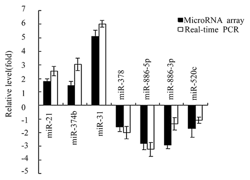
To determine whether miRNAs are involved in the development of the resistance to platinum in NSCLC cells, a comprehensive miRNA profiling of platinum-resistant lung cancer cell line A549/CDDP and its parental cell line A549 was performed. It was shown that 21 human miRNAs examined were deregulated more than 1.5-fold in A549 /CDDP cells compared with those in A549 cells, with 10 miRNAs upregulated and 11 miRNAs downregulated (average fold change were listed ). These miRNAs may play an important role in the development of platinum resistance in NSCLC cells. To verify the results obtained by microarray profiling, we selected seven differentially expressed miRNAs (miR-21, miR-31, miR-374b, miR-378, miR-886-5p, miR-886-3p, and miR-520c) at random for real-time PCR analysis in one pair of samples of the two cell lines. The analysis confirmed the results obtained by the microarray as shown in .
Table 2. List of deregulated miRNAs at > 1.5-fold in A549/CDDP vs. A549 cells
Role of miR-21 in platinum resistance in NSCLC
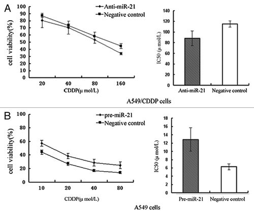
The reason to select miR-21 for further study is because previous studies have suggested that it regulates several important molecular behaviors of cancer, including modulation of the drug sensitivity of cells.Citation6,Citation7 To test whether miR-21 indeed has a function in platinum resistance in non-small cell lung cancer cells, gain-of-function and loss-of-function approaches in A549 and A549/CDDP cells were used, which express relatively high and low levels of miR-21, respectively. Zeiss fluorescence microscopy was used to determine the fluorescence signal, and the fluorescent carrier rate is approximately 80%. To further investigate the transfection efficiency, increased or decreased expression of miR-21 upon transfection was assessed by real-time PCR. The expression levels of miR-21 were increased or reduced nearly 10- to 20-fold change after the transfection of pre-miR-21 or anti-miR-21, respectively. MTT assay revealed that A549/CDDP cells transfected with a specific inhibitor of miR-21, which exhibited highly enhanced sensitivity to CDDP, compared with those transfected with scrambled oligonucleotides as indicated by significantly decreased IC50 values. Both miR-21 and the negative control survival were starting at 100%. The IC50 of A549/CDDP cells transfected with anti-miR-21 was 1.30 times lower (p < 0.05) compared with the A549/CDDP cells transfected with scrambled oligonucleotides (). On the other hand, overexpression of miR-21 level in A549 cells by transfecting the pre-miR-21 led to decreased sensitivity of A549 cells to CDDP. The IC50 of A549 cells transfected with pre-miR-21 was 2.04 times higher (p < 0.05) compared with the A549 cells transfected with scrambled oligonucleotides (). The above data indicate that modulation of miR-21 expression could alter the platinum sensitivity of lung cancer cells.
Figure 2. Effects of miR-21 on drug sensitivity of NSCLC cells. (A) Effects of anti-miR-21 on A549/CDDP cells. Transfection of the A549/CDDP cells with anti-miR-21 increases their sensitivity to platinum treatment. (B) Effects of pre-miR-21 on A549 cells. Transfection of the A549 cells with pre-miR-21 decreases their sensitivity to platinum treatment.

miR-21 expressions significantly increased in patients with chemotherapy-resistant NSCLC
To further verify miR-21 expression level associated with clinical characteristics including resistance to platinum based adjuvant chemotherapy in patients with NSCLC, tumor tissue specimens from 58 patients who were identified to fit study criteria were analyzed by miR-21 Taqman Real-time PCR. Correlation between miR-21 expression level and clinicopathological characteristics of NSCLC is summarized in . A statistically significant association between miR-21 expression level and sensitivity to platinum of NSCLC was observed in this study (p = 0.007). MiR-21 expression was indeed significantly increased in resistant response patients as compared with their counterparts with sensitivity response (median of miR-21 expression ratio = 5.798, p = 0.000).
Table 3. Comparison of several clinicopathologic factors and miR-21 expression level in 58 NSCLC patients undergone adjuvant platinum based chemotherapy
Higher expression of miR-21 significantly associated with shorter DFS of patients with NSCLC
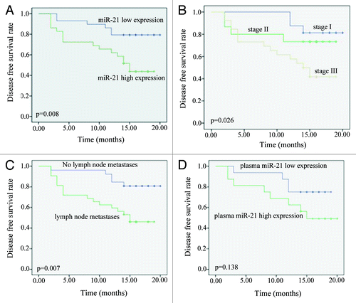
In this study, miR-21 was identified as a potential predictor for platinum based adjuvant chemotherapy resistance in patients with NSCLC. It was further investigated whether miR-21 could also serve as a prognostic marker for recurrence of the disease in patients with NSCLC received adjuvant platinum based treatment. Kaplan-Meier survival analysis indicated that higher expression of miR-21 (based on median level) was significantly associated with shorter DFS of the patients as compared with the lower miR-21 expression group (p = 0.008, n = 58; ). Interestingly, the Ct value of miR-21 was not detected in a patient, the only stage IV patient among the 58 patients, who were already alive more than 19 mo after the surgery with no recurrence (the RNU6B can be detected in this patient’s tissue RNA). Aside from miR-21 expression, DFS analysis of other clinicopathological factors also revealed that clinical TNM stage and lymph node metastases were associated with prognosis of the patients with NSCLC (). (n = 58, p = 0.026 and p = 0.007).
Figure 3. Kaplan-Meier estimation of Disease Free Survival time (DFS) of NSCLC. (A) DFS analysis of patients according to the miR-21 relative expression. The expression levels of miR-21 in 58 patients were measured by Real-time PCR. High expression is based on the median values of miR-21 relative expression. High miR-21 expression (p = 0.008) had a significant relationship with patient’s shorter DFS. (B) DFS analysis of patients according to their clinical stage. Patients were classified as either early or late clinical stage. Clinical stage had a significant relationship with patient’s DFS (p = 0.026). (C) DFS analysis of patients according to their lymph node status. Patients classified as either lymph node metastasis positive or negative. Lymph node metastasis was found to be strongly associated with poor outcome (p = 0.007). Log-rank p values are from Kaplan-Meier analysis. (D) DFS analysis of patients according to the miR-21 relative expression in plasma samples of 32 patients. The increased expression level of miR-21 in plasma is marginally associated with shorter DFS (p = 0.138).

Since lymph node metastasis and TNM stage are also related to DFS, we also evaluated the correlation between lymph node metastasis and miR-21 expression, and the correlation between TNM stage and miR-21 expression. Although lymph node metastasis group had a slight higher expression of miR-21 than that in negative group, but there was no significant difference (p > 0.05; Mann-Whitney test). Moreover, there was no statistical difference in the miR-21 expression among TNM I, II and III groups (p > 0.05; Kruskal-Wallis test). These data was consistent with the data in about the correlation between clinicopathological factors and miR-21 expression.
Univariate Cox proportional hazard regression analysis revealed that high miR-21 expression was the effective predictive factor for lower DFS of patients with NSCLC (p = 0.014, Hazard ratio [HR] = 3.625, 95% confidence interval [CI]:1.276–8.357). Other clinicopathological parameters, including clinical stage (p = 0.015, HR = 2.090, 95% CI: 1.153–3.791) and lymph nodes status (p = 0.013, HR = 3.532, 95% CI: 1.300 -9.595), were also found to be predictive factors for poor prognosis of NSCLC patients (). Moreover, multivariate Cox proportional hazard regression analysis revealed that high-level miR-21 expression (p = 0.032, HR = 2.820, 95% CI: 1.091–7.285) was also the most significantly unfavorable prognostic factor independent of other clinicopathological factors.
Table 4. Univariate and multivariate analysis for factors related to DFS using the COX proportional hazard model
Relationship between the miRNAs in plasma and primary lung cancer tissues
Among the 58 patients with lung tumor tissue samples, 32 patients also had the plasma specimen. Real time PCR of miR-21 was conducted in plasma to compare the expression level with that in their corresponding tumor tissues. The data showed that primary lung cancer tissues and paired plasma samples from 22 patients (22/32 = 68.75%) had similar tendencies concerning the miR-21 levels, which indicated a positive correlation between miR-21 expression detected in plasma samples and the expression detected in the corresponding tumor tissue samples (p = 0.034; correlation coefficient, 0.375, ). Interestingly, 5 (15.63%) patients with higher miR-21 expression in plasma had lower mir-21 expression in the corresponding primary tumors. Similarly, five patients with higher miR-21 expression in tumor had lower miR-21 expression in the corresponding plasma samples. These above data indicated that the level of plasma miRNAs might reflect the expression level of tumor miRNAs. However, the increased expression level of miR-21 in plasma is only marginally associated with the poor response to the platinum (p = 0.186) and DFS (p = 0.138, ) in these 32 patients.
Table 5. Correlation of miR-21 expression levels between plasma samples and corresponding primary tumor tissue samples
miR-21 regulated platinum resistance in lung cancer cells which is associated with the change of PTEN and Bcl-2 expression
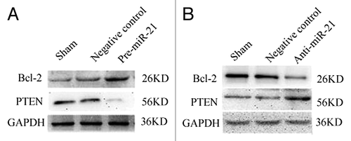
To dissect the molecular mechanism underlying the miR-21-associated alteration of the sensitivity of platinum in lung cancer cell lines, western blot analysis for the potential miR-21 related proteins were conducted, which may be involved in the drug resistance. Therefore, it was tested whether miR-21 regulated the platinum sensitivity in lung cell growth by triggering PTEN or apoptosis pathways as suggested by previous studies. A549/CDDP and A549 cells were transfected with either the anti-miR-21 or pre-mir-21 and scrambled oligonucleotides, and the protein levels of PTEN and Bcl-2 were determined 72 h after transfection. Our data shows that transfection of A549 cells with pre-miR-21 resulted in a decrease of PTEN levels and an increase of Bcl-2 (), whereas transfection of A549/CDDP cells with anti-miR-21 resulted in an increase of PTEN levels and a decrease of Bcl-2 ().
Figure 4. Expression of PTEN and BCL-2 in the A549 and A549/CDDP cells assessed by western blot after transfected with either scrambled or pre-miR-21 and anti-miR -21 expression. (A) The effects of miR-21 on the expression of PTEN and BCL-2 in A549 cells. Transfection of A549 cells with pre-miR-21 resulted in a decrease of PTEN levels and an increase of Bcl-2. (B) The effects of miR-21 on the expression of PTEN and BCL-2 in A549/CDDP cells. Transfection of A549/CDDP cells with anti-miR-21 resulted in an increase of PTEN levels and a decrease of Bcl-2. Sham: A549 or A549/CDDP without transfection; Negative control: A549 or A549/CDDP with transfection of Anti-miR and Pre-miR.

Discussion
NSCLC continues to be the leading cause of cancer mortality worldwide. It is still a major hurdle that cancer cells become resistant to chemotherapeutic agents during treatment. At present, the resistance to platinum is considered as a multifactorial phenomenon involving several major cellular mechanisms. However, the molecular mechanisms of its resistance remain to be identified. Additionally, biomarkers at the mRNA and protein levels have been widely studied to explore mechanisms that determine the pharmacological response, whereas levels of mRNA and the encoded proteins are often not proportional, which could be considered to be partly due to the regulatory influences of miRNAs. Furthermore, miRNA signatures might be more effective than mRNA signatures in categorizing, detecting, and predicting the course of human cancers as well as in characterizing developmental origins of tumors.Citation9 Despite the well-established role of miRNA in cancer, the role of miRNA in cancer drug resistance remains largely unexplored.
This study identified the miRNA signature associated with platinum resistance in lung cancer cells by using miRNA microarray on the A549 and A549/CDDP. The A549/CDDP cell line is a cell model which is most commonly used to study platinum-resistant of lung cancer.Citation10,Citation11 Since resistant cell line and its parental cell line are homologous, they are often used in the comparative studies for drug resistance.Citation12-Citation14 Among the profile generated by miRNA microarray, miR-21, with well reported oncogenic activity in various cancers, is highly expressed in the platinum-resistant A549/CDDP cell line compared with its parent A549 cell line. This data is consistent with our previous report,Citation15 which states that overexpressed miR-21 is associated with the poor overall post-operative survival of patients with NSCLC. In general, among the clinical marker candidates, physicians favor the markers which are upregulated in carcinoma samples and functional studied. Despite the well-established role of miR-21 in cancer development and progression, the role of miR-21 in NSCLC drug resistance remains largely unclear. Moreover, in this study, the data shows that knockdown of miR-21 overrides platinum resistance in A549/CDDP cells, whereas ectopic expression of miR-21 resulted in the decreased sensitivity of A549 cells to platinum. This result is consistent with a previous in vitro study,Citation16 which suggested that miR-21 levels could play a substantial role in cancer cells’ sensitivity to chemotherapeutic agents. In view of these findings, miR-21 was selected in this study for further investigation.
To confirm the correlation between miR-21 expression and the response to chemotherapy in patients with NSCLC, we selected the clinical samples from patients who had received platinum based combination chemotherapy. Tissue samples were analyzed by miRNA Taqman Real-time PCR analysis. In the present study, we observed a robust association with miR-21 expression level and NSCLC therapeutic outcome. MiR-21 was found to have significantly higher expression levels in the groups of patients that were resistant as compared with sensitivity to treatment by platinum based adjuvant chemotherapy. Additionally, patients with lower miR-21 expression had significantly longer DFS times after adjuvant platinum-based chemotherapy treatment, which suggests that these patients might have benefited from the treatment. It is to be noted that the Ct value of miR-21 was not detected in a patient, the only stage IV patient among the 58 patients, who were already alive more than 19 mo after the surgery with no recurrence. Since lymph node metastasis and TNM stage is also related to DFS, we evaluated both the correlation between lymph node metastasis and miR-21 expression, and the correlation between TNM stage and miR-21 expression. Although high expression of miR-21 was reported associated with lymph node metastasis or clinical stage in some cancers, such as breast cancer,Citation17,Citation18 esophageal carcinoma,Citation19 in some other cancers, such as pancreatic cancer,Citation20 colon cancer,Citation21 miR-21 expression is not associated with lymph node metastasis. In the present study, there was no statistical difference in the miR-21 expression between lymph node positive and negative groups, and among TNM groups (p > 0.05). These data was consistent with both our previously published workCitation15 and other’s study.Citation22 Moreover, the Cox proportional hazard regression model in the present study showed that miR-21 expression levels were reversely related to poor DFS, independent of other clinical covariates. Our results suggest that miR-21 expression may be a useful prediction indicator, in addition to other clinical parameters, to help identify patients at a higher risk of terminal cancer.
In the year 2008, two research groupsCitation23,Citation24 had first reported that serum and plasma contain a large amount of stable miRNAs and that the expression of these miRNAs show great promise as novel non-invasive biomarkers for the early diagnosis of various cancers and other diseases.Citation25-Citation39 These researchers also showed the high stability of circulating miRNAs after RNase digestion and other harsh conditions such as boiling, extremes of pH, extended storage, and freeze-thaw cycles. Thus, the high stability characteristics of circulating miRNAs would potentially provide advantages as biomarkers when compared with those of other nucleic acids, such as circulating DNA and mRNA. However, it remains unclear whether these circulating miRNAs are necessarily tumor-derived and how the miRNAs are protected from degradation or how they get into the blood. It may be due to dead tumor cells being lysed or tumor cells releasing miRNAs into their surroundings.Citation23,Citation24 Furthermore, it has been recently debated whether dysregulation of miRNA in peripheral blood is consistent with that in the matched tumor tissue like other molecular markers in peripheral blood.Citation27,Citation29 These findings led to the investigation of the significance of plasma miR-21 in patients with NSCLC who received systemic platinum based chemotherapy. In the present study, to investigate the relationship between the miRNA abundances in plasma and primary lung cancer tissues, preoperative plasma samples were chosen because patients was in tumor-bearing status before surgery.Citation25,Citation28 Moreover, they have not received any therapeutic intervention and this point would actually be more conducive to the clinical analysis. The comparison between expressions of miR-21 in plasma and the corresponding tumor tissue demonstrated that plasma and primary NSCLC tumor tissue samples showed similar patterns regards to the abundance of miR-21 in 68.75% cases. Therefore, the results of this study demonstrated the possibility of using plasma as a surrogate tissue sample to measure miRNA expression. Additionally, within the author’s knowledge, this is the first description of the comparison between expressions of miRNA in matched lung cancer tissues and plasma in a cohort in the treatment efficacy study of NSCLC. A close correlation between circulating miRNAs and tumor miRNAs has also been observed in previous published studies that had smaller sample sizes (only four paired tumor and plasma sample from lung adenocarcinoma cases).Citation29 However, it should be noted that a higher miR-21 expression was found only in either the plasma samples or the tumor samples in nearly 30% of the patients. One possible explanation for these discrepancies remain, is the heterogeneity of genetic abnormalities in the primary tumors. The dilution of miRNA derived from noncancerous tissues, such as inflamed tissues, might impede the detection of miRNA in serum, despite the presence of this miRNA in tumors.Citation30 Moreover; the source of the miRNA in circulation is complex. In addition to the secretion mechanism of tumor cell lysis like most literature showed, other mechanisms maybe also involved, such as host immune system or stress response.Citation31 Nevertheless, the inability to obtain primary tumor tissues, particularly through repeated biopsies from patients with NSCLC makes the use of plasma as a surrogate tissue for prognostic and predictive related miRNA analysis clinically important.
Consistent with tumor tissue sample, plasma miR-21 was not related to clinical factors including TNM stage, histological classification, and lymph node status. However, it should be noted that our previous study tested plasma miR-21 expression in 63 patients with stage I-IV NSCLC (more than half patients are at stage IV). The data indicated that plasma miR21 expression was significantly higher in stage III-IV patients than in stage I–II patients (p = 0.043).Citation32 This difference between our present and previous study could be mainly due to two reasons. First, the present study analyzed the stage I–III only patients’ plasma miR-21-TNM stage; however our previous study chose I–IV patients which have more than half cases in stage IV. Besides the impact of weight of every stage percentage to statistical analysis, we hypothesized that plasma miR-21 may play a more important role in the progression of advanced NSCLC especially in IV stage patients. Second, in the present study, plasma samples were collected prior to definitive surgical management. However, in our previous study, blood samples were taken before chemotherapy in both operable and non-operable patients (in operable patients: post-operation blood samples were collected). Some published data indicated that surgery can decrease miRNA expression. Liu et al.Citation50 showed that plasma miR-31 in oral squamous cell carcinoma (OSCC) patients was remarkably reduced after tumor resection suggesting that this marker is tumor associated.Citation33 Wong et al.Citation51 showed that the plasma miR-184 levels were 10-fold reduced after the surgical removal of tongue SCC tumor. Thus, they also indicated that the higher plasma miRNA levels were due to absorption of the miRNA into the circulation from a large number of cancer cells with highly expressed miR-184 in the body before surgery. The plasma level therefore reduced significantly after the surgical removal of the tumor.Citation34 Thus, Different plasma sample collection time point between our present and previous study may alsoresult to the different clinical analysis result. Nevertheless, a larger scaled study to confirm our data is required in the future.
Although plasma and primary tumor tissue samples showed similar miR-21 expression tendencies, the correlation between plasma miR-21 level and response to the platinum based adjuvant chemotherapy and DFS is only marginally significant, which may be due to the modest sample size, since only 12 out of 32 developed resistance. Our previous study analyzed plasma miR-21 expression in 35 advanced (IV stage) NSCLC patients who had undergone 2 to 3 cycles of platinum based chemotherapy. And the results showed that plasma miR-21 expression was significantly higher in the progressive disease (PD) plus stable disease (SD) group than in the partial remission (PR) group (p = 0.049), suggesting that plasma miR-21 expression may correlate with sensitivity to chemotherapy in advanced inoperable NSCLC.Citation32 However, there are multiple differences between operable NSCLC and advanced NSCLC, such as the chemotherapy (adjuvant chemotherapy or advanced chemotherapy),Citation33-Citation35 patients’ TNM stage, calculation methods of response definition, treatment invention before chemotherapy (focal resection or not), tumor burden, and miRNA test methodsCitation36,Citation37 in these two studies. Thus the authors do not think that these two studies can provide significant cross-reference to each other. Nevertheless, whether plasma miR-21 could be used as sensitive and specific surrogate tumor markers for prediction platinum based adjuvant chemotherapy in NSCLC requires further studies in serially taken blood samples of larger cohort of patients with long-term follow-up.
The exact molecular mechanism of miR-21 dysregulation in the acquisition of cancer cell resistance to platinum is currently unclear. It may be related to multi-level regulatory control such as anti-apoptosis, proliferation and invasion-related genes. MiR-21 has been shown to play critical roles in cancer biology including chemoresistance in cholangiocarcinoma cells and breast cells. Upregulation of miR-21 induces cholangiocarcinoma cell survival and gemcitabine resistance primarily through targeting the PTEN dependent PI3K/Akt pathway.Citation6 Inhibition of miR-21 was shown to increase the sensitivity to topotecan in breast cancer cells partly by regulating BCL2 induced anti-apoptosis indirectly in MCF-7 cells.Citation7 Our further investigation using cancer cell lines confirmed that miR-21 is functionally involved in lung cancer cells response to platinum partly by the potential effect of PTEN and Bcl-2, which is consistent with previous data. Furthermore, it has been well documented that both constitutive deregulation of PTEN and upregulation of Bcl-2 contribute to platinum resistance in different types of tumors, including NSCLC. Loss of functional PTEN leads to increased activity of AKT and the mammalian target of rapamycin (mTOR) kinase pathways, which can promote both cell survival and proliferation through phosphorylation and inactivation of several downstream mediators.Citation38 The antiapoptotic protein Bcl-2 contributes to a more chemoresistant phenotype of lung cancer cell to ciaplatin, but not to docetaxel.Citation39 However, in contrast to miR-21 directly targeting PTEN in holangiocarcinoma cell,Citation6 Si et al. reported that miR-21 may indirectly regulate Bcl-2 expression as shown in a breast cancer study.Citation7 Their research was suggested that anti-miR-21 suppresses expression of a gene(s) that negatively regulates Bcl-2 expression. Our present study, consistent with those above researches, showed that transfection of A549/CDDP cells with anti-miR-21 increased the expression of the tumor suppressor-PTEN protein and decreased the anti-apoptosis factor-Bcl-2 protein. In contrast, the pre-miR-21 decreased PTEN expression and increased Bcl-2 expression in the parental cells A549. Thus, the data in our study indicates that miR-21 could play a role in the resistance of NSCLC cells to platinum in part due to regulation of some well-known genes such as PTEN and Bcl-2.
Consistent with many studies focused on the function of miRNAs, this study has only analyzed two potential target proteins. However, cancer drug resistance is considered as a multifactorial phenomenon involving several major mechanisms, such as increased repair of DNA damage, defective DNA damage response, reduced apoptosis, altered metabolism of drugs, increased energy-dependent efflux of chemotherapeutic drugs and disregulated chemoresistance related proteins like multidrug resistance proteins (MDRs) that diminish the ability of cytotoxic agents to kill cancer cell.Citation3,Citation40 For instance, xenobiotic enzyme and its related pathway also have been reported to affect chemoresistance in human lung adenocarcinoma cells.Citation41 In addition, recently a prevailing hypothesis states that cancer stem cells (CSCs) persist in tumors causing relapse after chemotherapy, thus rendering these cells as critical targets responsible for tumor resistance and recurrence.Citation42,Citation43 A study by Bao et al. showed that CDF (a novel synthetic compound) could significantly inhibit the sphere-forming ability (pancreatospheres) of pancreatic cancer (PC) cells consistent with increased disintegration of pancreatospheres, which was associated with attenuation of CSC markers (CD44 and EpCAM), especially in gemcitabine-resistant (MIAPaCa-2) PC cells containing high proportion of CSCs consistent with increased miR-21 and decreased miR-200. Their results strongly suggest that the anti-tumor activity of CDF is associated with inhibition of CSC function via downregulation of CSC-associated signaling pathways.Citation44 Another study by Misawa et al. defined the critical role of miR-21 in maintenance of the chemoresistant phenotype of SP cells. Targeting miR-21 may provide a new strategy for cancer therapy by impairing resistance to chemotherapy in CSCs.Citation45 Thus, we speculate that miR-21 may be related to multi-level regulatory control as above mentioned pathways. Chemo resistant-related miR-21-targets are definitely not just one or two, but rather a series of factors. Moreover, since miRNA regulates the target gene expression either by translational suppression or by mRNA destabilization of mRNAs with imperfect complementary sequences, common in mammals,Citation46 attention on transcriptional level of these targets is also essential. Therefore, further study focused on more targets and their regulatory network is required in the near future.
In summary, although there are some limitations in the present study, such as modest sample size, lacking of overall survival data and failure in including multiple targets and deep functional investigation, this study still provides important evidence that miRNAs, in particular, miR-21 is critical in platinum resistance to human NSCLC cell. Patients with complete NSCLC rectomy and lower expression of miR-21 tumors derived a substantial benefit from adjuvant platinum-based chemotherapy, as compared with patients with higher miR-21 expression tumors. The data also suggested that circulating plasma miRNA may be used as a surrogate tissue for miR-21 expression analysis in NSCLC. Further investigation indicated that miR-21 could modulate the sensitivity of NSCLC cells to platinum, at least in part, by regulating PTEN and Bcl-2 expressions. This study translates to a valuable pilot study demonstrating the potential usefulness of NSCLC tissue or circulating miR-21 as a candidate biomarker combined with traditional factors for further prospective prediction of the clinical benefits from platinum based adjuvant chemotherapy and to monitor therapeutic plans. Additionally, miR-21 could also serve as a potential target for chemosensitizing strategies. Clinical studies with specifically defined treatment regimens, including more miR-21-targets and larger sample sizes are necessary. Further studies focused on the mechanism of plasma miRNAs generation and physiological function are also urgently needed. In addition, the role of miR-21 in platinum based chemoresistance warrants further in vitro and in vivo studies.
Materials and Methods
Cell lines and cultures
The human lung adenocarcinoma cell line A549 and the resistant cell line A549/CDDP were purchased from Mshar Bioscience and Technology. The cells were cultured in RPMI-1640 medium (Hyclone) supplemented with 10% fetal calf serum (Hyclone) at 37°C in a humidified atmosphere containing 5% CO2. To maintain the drug resistance, platinum (with final concentration of 1 μg/ml) was added to the culture media for A549/CDDP cells.
Clinical samples
In total, 58 snap-frozen NSCLC tissue samples were evaluated for miRNA expression. These samples were collected from patients in the department of Cardiothoracic Surgery, the First Affiliated Hospital of Nanjing Medical University and the department of thoracic Surgery, Jiangsu Cancer Hospital between January 2009 and October 2009. All tissue samples were from primary sites, and were immediately snap-frozen and stored in liquid nitrogen. This study was approved by the Institutional Review Boards of the First Affiliated Hospital of Nanjing Medical University. All cases were histopathologically diagnosed as having NSCLC without radiotherapy or chemotherapy before surgical operation. Moreover, all cases received at least 2–4 cycles of the platinum-based chemotherapy as the first-line adjuvant therapy after surgical operation. The clinical and histological diagnosis of tumors was based on criteria of the American Joint Committee on Cancer/International Union against Cancer staging system (AJCC/UICC) and the TNM stage was determined according to criteria revised in 2009. Samples chosen for analysis contained > 60–70% tumor cells. Patients suffered with recurrent or progressive disease during first line platinum based chemotherapy or within 12 mo post-completion of first line therapy were considered as platinum-resistant. Patients with no recurrence or with recurrences beyond 12 mo were defined as platinum sensitive. Disease free survival time (DFS) was calculated as the time from the date of surgery to the time of detected recurrence/progression or death from any cause.Citation47 The final follow-up was in November, 2010.
Of the 58 patients’ cancer samples, 32 had matching blood samples. Up to 5 mL of blood from each patient was collected in an EDTA tube. Each blood sample was centrifuged at 2,000 g for 20min at 4°C to spin down the blood cells, and then the supernatant plasma was carefully transferred into an RNase-free microcentrifuge tube for extraction of RNA. All blood samples were processed and plasma was frozen within 4 h of the blood draw.
RNA isolation
Total RNA was isolated from 100 to 500 mg of frozen tissue or 106 to 107 cultured cells with mirVanaTM miRNA isolation kit (Ambion 1560). While Total RNA was isolated from 400 µL plasma using the mirVana PARIS kit (Ambion) following the manufacturer’s protocol for liquid samples (Ambion), modified such that samples were extracted twice with an equal volume of acid-phenol chloroform (supplied by the Ambion kit).Citation23 RNA concentration was assessed by measuring absorption (A260/A280) on the NanoDrop Spectrophotometer ND-1000 (NanoDrop).
MiRNA microarray and analysis
Prior to experimentation, A549/CDDP cells were cultured for 1 week without platinum. The isolated RNAs from the A549 and A549/CDDP cell lines were then labeled by using the miRCURY Hy3/Hy5 Power labeling kit and hybridized respectively on the miRCURY LNA Array (v.11.0) (Exiqon). The miRCURY LNA Array (v.11.0) covers miRNAs annotated in Sanger miRBase version 11.0, which contains more than 2,056 capture probes, including 847 human miRNA genes. Three independent hybridizations were performed on three chips for each sample, while every miRNA was plated in quadruplicate on each chip. Microarray images were acquired using a Genepix 4000B scanner (Axon Instruments) and were processed and analyzed with Genepix Pro 6.0 software (Axon Instruments) and Excel. The ratio of red signal to green signal was calculated after background subtraction and normalization using the global Lowess (Locally Weighted Scatter plot Smoothing) regression algorithm (MIDAS, TIGR Microarray Data Analysis System), which produces the best within-slide normalization to minimize the intensity-dependent differences between the dyes. The positive effect of this normalization is illustrated in two different plots for each slide. To reduce between-slide variability, normalization was performed by scale normalization. Replicated spots on the same slide have been averaged by getting a median ratio of replicated spots.
Validation of miRNA microarray results
To warrant the accuracy of the microarray analysis, real-time PCR validation with the same RNA preparations used in microarray analysis was performed (total RNA was prepared from one pair of specimens). First, CDNA was prepared by miRNA-specific primer (miR-21, miR-31, miR-374b, miR-378, miR-886-5p, miR-886-3p, miR-520c)Citation17 and then, real-time quantification RT-PCR for these miRNAs and an endogenous control (RNU6B) were performed on an ABI PRISM 7900 system (Applied Biosystems). These miRNA-specific RT primers were designed by primer premier 5.0, and are summarized in .
Table 1. RT primers for amplification of the human mature miRNAs used in validation of microRNA microarray results
Stem-loop real-time reverse transcription-PCR (TaqMan miRNA assay)
Expression of mature miRNAs was analyzed by TaqMan miRNA Assay (Applied Biosystems) in accordance with the manufacturer's instructions. Briefly, single-stranded cDNA was synthesized from 10 ng of total RNA in a 15 µL reaction volume using the TaqMan MiRNA Reverse Transcription kit (Applied Biosystems). The reactions were incubated on a GeneAmp PCR System (Bio-Rad) for 30 min at 16°C, 30 min at 42°C, 5 min at 85°C, and then held at 4°C. All reverse transcriptions and no-template controls were run at the same time. PCR amplification was performed using sequence-specific primers on the Applied Biosystems 7500 real-time PCR system (Applied Biosystems). The 20 μL PCR reactions included 10 μL of 2 × Universal PCR Master Mix (no AmpErase UNG), 1 μL of each 10 × TaqMan MiRNA Assay Mix and 1.33 μL of reverse transcription product. The reactions were incubated in a 96-well plate at 95°C for 10 min, followed by 40 cycles at 95°C for 15 sec and 60°C for 1 min. Normalization was performed with the small nuclear RNA U6 (RNU6B; Applied Biosystems). RNU6B was also identified using the TaqMan RNU6B assay for normalizing the levels of miR-21.Citation48 Each sample that didn’t have a template was analyzed in triplicate. Data was analyzed using SDS software, version 1.4. Relative expression was calculated using the comparative Ct method.Citation49
MiRNA transfection
All miRNA precursors, antisense inhibitors or controls were purchased from Ambion. The A549/CDDP cells were seeded in 6-well plates (2 × 10⁵ cells/well) in OPTI-MEM. OPTI-MEM is a serum-free medium without antibiotics (Gibco/BRL). A549/CDDP cells were then transfected with scrambled RNA oligonucleotide (anti-miRNA negative control: A carboxyfluorescein (FAM)-labeled RNA oligonucleotide designed for monitoring uptake of Anti-miR miRNA Inhibitors by fluorescence microscopy or other fluorescence-based technique) or 50 nmol/L anti-miR-21 in three independent replicates using X-tremeGENE siRNA Transfection Reagent (Roche Applied) in accordance with the manufacturer’s protocol. Similarly, The A549 Cells were also plated in 6-well plates (2 × 10⁵ cells/well) in OPTI-MEN and transfected with scrambled RNA oligonucleotide (pre-miRNA negative control: A carboxyfluorescein (FAM)-labeled RNA oligonucleotide designed for monitoring uptake of pre-miRNA by fluorescence microscopy or other fluorescence-based technique) or 50 nmol/L Pre-miR-21 in three independent replicates using X-tremeGENE siRNA Transfection Reagent (Roche) in accordance with the manufacturers’ procedures. Six to eight hours after transfection, Zeiss fluorescence microscopy and Realtime PCR were used to determine the transfection efficiency. RNA and protein were extracted after 24 and 72 h of transfection respectively. RNA was used for transfection validation and protein was used for western blot analysis.
In vitro drug sensitivity assay
About 48 h after transfection, cells were reseeded in 96-well plates in the presence of miRNA at density of 1 × 10⁴ per well and treated with Platinum at a concentration range of 5–320 μmol/L in the culture medium for another 48 h. For the analysis of the effects of precursors or inhibitor of miR-21 on drug sensitivity, MTT (methyl thiazolyl tetrazolium) assay was performed 48 h post-drug addiction. The concentration at which each drug produced 50% inhibition of growth (IC50) was estimated by the relative survival curve.
Western blot analysis
Total proteins were extracted using Total protein extraction kit (Keygen) following the manufacturer's protocol. The protein samples (50 µg) were then subjected to 12 or 10% SDS-PAGE and transferred to PVDF (Roche Diagnostics) membrane. Membranes were blocked at room temperature for 2 h with 5% nonfat powered milk in Tris-buffered saline containing 0.05% Tween 20, then incubated for 45 min with primary antibody, washed and incubated with secondary antibody, and visualized by chemi-luminescence. The chemi-luminescence signal was imaged using a ChemiDoc XRS (Bio-Rad) and quantified using Quantity One software (Bio-Rad). The primary antibodies used were: Anti-Bcl-2 (1:500, Bioworld), anti-PTEN (1:500, Bioworld) and GAPDH (1:1000, Santa Cruz). Horseradish peroxidase (HRP) conjugates of goat anti-rabbit IgG and goat anti-mouse IgG (1:2000, Zhongshan Biotech) were used as the secondary antibody.
Statistical analysis
Relationships among miR-21 and various clinicopathological factors were analyzed by the Chi square test. Expression levels between groups of samples were compared using the Mann-Whitney non-parametric test. Kaplan-Meier curves were used to estimate DFS and were compared with the use of log rank statistics. The joint effect of covariables was examined using the Cox proportional hazard regression model. These above tests were two-tailed, and the significance level was set at p < 0.05.
Acknowledgments
This study was supported by the Natural Science Foundation of Jiangsu Province (BK2010578) and the National Natural Science Foundation of China (Grant No. 81101759).
Disclosure of Potential Conflicts of Interest
No potential conflicts of interest were disclosed.
References
- Shen HB, Yu SZ. Epidemiological status of lung cancer in China and strategies for prevention. Bulletin Chinese Cancer 2004; 5:283 - 5
- Gurubhagavatula S, Lynch TJ. The role of adjuvant chemotherapy for non-small cell lung cancer. Semin Respir Crit Care Med 2005; 26:298 - 303; http://dx.doi.org/10.1055/s-2005-871988; PMID: 16052431
- Sève P, Dumontet C. Chemoresistance in non-small cell lung cancer. Curr Med Chem Anticancer Agents 2005; 5:73 - 88; http://dx.doi.org/10.2174/1568011053352604; PMID: 15720263
- Yin M, Yan J, Voutsina A, Tibaldi C, Christiani DC, Heist RS, et al. No evidence of an association of ERCC1 and ERCC2 polymorphisms with clinical outcomes of platinum-based chemotherapies in non-small cell lung cancer: A meta-analysis. Lung Cancer 2011; In press PMID: 21075476
- Ruan K, Fang X, Ouyang G. MicroRNAs: novel regulators in the hallmarks of human cancer. Cancer Lett 2009; 285:116 - 26; http://dx.doi.org/10.1016/j.canlet.2009.04.031; PMID: 19464788
- Meng F, Henson R, Lang M, Wehbe H, Maheshwari S, Mendell JT, et al. Involvement of human micro-RNA in growth and response to chemotherapy in human cholangiocarcinoma cell lines. Gastroenterology 2006; 130:2113 - 29; http://dx.doi.org/10.1053/j.gastro.2006.02.057; PMID: 16762633
- Si ML, Zhu S, Wu H, Lu Z, Wu F, Mo YY. miR-21-mediated tumor growth. Oncogene 2007; 26:2799 - 803; http://dx.doi.org/10.1038/sj.onc.1210083; PMID: 17072344
- Yang H, Kong W, He L, Zhao JJ, O'Donnell JD, Wang J, et al. MicroRNA expression profiling in human ovarian cancer: miR-214 induces cell survival and cisplatin resistance by targeting PTEN. Cancer Res 2008; 68:425 - 33; http://dx.doi.org/10.1158/0008-5472.CAN-07-2488; PMID: 18199536
- Lu J, Getz G, Miska EA, Alvarez-Saavedra E, Lamb J, Peck D, et al. MicroRNA expression profiles classify human cancers. Nature 2005; 435:834 - 8; http://dx.doi.org/10.1038/nature03702; PMID: 15944708
- Liu LZ, Zhou XD, Qian G, Shi X, Fang J, Jiang B. HAKT1 amplification regulates cisplatin resistance in human lung cancer cells through the mammalian target of rapamycin/p70S6K1 pathway. Cancer Res 2007; 67:6325 - 32; http://dx.doi.org/10.1158/0008-5472.CAN-06-4261; PMID: 17616691
- Li JJ, Ding Y, Li DD, Peng RQ, Feng GK, Zeng YX, et al. The overexpression of ERCC-1 is involved in the resistance of lung cancer cells to cetuximab combined with DDP. Cancer Biol Ther 2009; 8:1914 - 21; http://dx.doi.org/10.4161/cbt.8.20.9439; PMID: 20009541
- Akao Y, Noguchi S, Iio A, Kojima K, Takagi T, Naoe T. Dysregulation of microRNA-34a expression causes drug-resistance to 5-FU in human colon cancer DLD-1 cells. Cancer Lett 2011; 300:197 - 204; http://dx.doi.org/10.1016/j.canlet.2010.10.006; PMID: 21067862
- Xia L, Zhang D, Du R, Pan Y, Zhao L, Sun S, et al. miR-15b and miR-16 modulate multidrug resistance by targeting BCL2 in human gastric cancer cells. Int J Cancer 2008; 123:372 - 9; http://dx.doi.org/10.1002/ijc.23501; PMID: 18449891
- Kovalchuk O, Filkowski J, Meservy J, Ilnytskyy Y, Tryndyak VP, Chekhun VF, et al. Involvement of microRNA-451 in resistance of the MCF-7 breast cancer cells to chemotherapeutic drug doxorubicin. Mol Cancer Ther 2008; 7:2152 - 9; http://dx.doi.org/10.1158/1535-7163.MCT-08-0021; PMID: 18645025
- Gao W, Yu Y, Cao HL, Shen H, Li XD, Pan SY. Deregulated expression of miR-21, miR-143 and s miR-181a in non small cell lung cancer is related to clinicopathologic characteristics or patient prognosis. Biomed Pharmacother 2010; 64:399 - 408; http://dx.doi.org/10.1016/j.biopha.2010.01.018; PMID: 20363096
- Blower PE, Chung JH, Verducci JS, Lin S, Park JK, Dai Z, et al. MicroRNAs modulate the chemosensitivity of tumor cells. Mol Cancer Ther 2008; 7:1 - 9; http://dx.doi.org/10.1158/1535-7163.MCT-07-0573; PMID: 18187804
- Yan LX, Huang XF, Shao Q, Huang MY, Deng L, Wu QL, et al. MiRNA miR-21 overexpression in human breast cancer is associated with advanced clinical stage, lymph node metastasis and patient poor prognosis. RNA 2008; 14:2348 - 60; http://dx.doi.org/10.1261/rna.1034808; PMID: 18812439
- Huang GL, Zhang XH, Guo GL, Huang KT, Yang KY, Shen X, et al. Clinical significance of miR-21 expression in breast cancer: SYBR-Green I-based real-time RT-PCR study of invasive ductal carcinoma. Oncol Rep 2009; 21:673 - 9; PMID: 19212625
- Hummel R, Hussey DJ, Michael MZ, Haier J, Bruewer M, Senninger N. MiRNAs and their association with locoregional staging and survival following surgery for esophageal carcinoma. Ann Surg Oncol 2011; 18:253 - 60; http://dx.doi.org/10.1245/s10434-010-1213-y; PMID: 20628822
- Dillhoff M, Liu J, Frankel W, Croce C. Bloomston. MMicroRNA-21 is overexpressed in pancreatic cancer and a potential predictor of survival. J Gastrointest Surg 2008; 12:2171 - 6; http://dx.doi.org/10.1007/s11605-008-0584-x; PMID: 18642050
- Shibuya H, Iinuma H, Shimada R, Horiuchi A, Watanabe T. Clinicopathological and Prognostic Value of MicroRNA-21 and MicroRNA-155 in Colorectal Cancer. Oncology 2010; 79:313 - 20; http://dx.doi.org/10.1159/000323283; PMID: 21412018
- Markou A, Tsaroucha EG, Kaklamanis L, Fotinou M, Georgoulias V, Lianidou ES. Prognostic value of mature microRNA-21 and microRNA-205 overexpression in non-small cell lung cancer by quantitative real-time RT-PCR. Clin Chem 2008; 54:1696 - 704; http://dx.doi.org/10.1373/clinchem.2007.101741; PMID: 18719201
- Mitchell PS, Parkin RK, Kroh EM, Fritz BR, Wyman SK, Pogosova-Agadjanyan EL, et al. Circulating microRNAs as stableblood-based markers for cancer detection. Proc Natl Acad Sci USA 2008; 105:10513 - 8; http://dx.doi.org/10.1073/pnas.0804549105; PMID: 18663219
- Chen X, Ba Y, Ma L, Cai X, Yin Y, Wang K, et al. Characterizationof miRNAs in serum: a novel classof biomarkers for diagnosis of cancer and other diseases. Cell Res 2008; 18:997 - 1006; http://dx.doi.org/10.1038/cr.2008.282; PMID: 18766170
- Huang Z, Huang D, Ni SJ, Peng ZL, Sheng WQ, Du X. Plasma microRNAs are promising novel biomarkers for early detection of colorectal cancer. Int J Cancer 2010; 127:118 - 26; http://dx.doi.org/10.1002/ijc.25007; PMID: 19876917
- Slaby O, Svoboda M, Michalek J. Vyzula RMicroRNAs in colorectal cancer: translation of molecular biology into clinical application. Mol Cancer 2009; 8:102; http://dx.doi.org/10.1186/1476-4598-8-102; PMID: 19912656
- Zhu W, Qin W, Atasoy U, Sauter ER. Circulating microRNAs in breast cancer and healthy subjects. BMC Research Notes 2009; 2:89; http://dx.doi.org/10.1186/1756-0500-2-89; PMID: 19454029
- Hu Z, Chen X, Zhao Y, Tian T, Jin G, Shu Y, et al. Serum MicroRNA Signatures Identified in a Genome-Wide Serum MicroRNA Expression Profiling Predict Survival of Non–Small-Cell Lung Cancer. J Clin Oncol 2010; 28:1721 - 6; http://dx.doi.org/10.1200/JCO.2009.24.9342; PMID: 20194856
- Rabinowits G, Gerçel-Taylor C, Day JM, Taylor DD, Kloecker GH. Exosomal microRNA: a diagnostic marker for lung cancer. Clin Lung Cancer 2009; 10:42 - 6; http://dx.doi.org/10.3816/CLC.2009.n.006; PMID: 19289371
- Bai H, Mao L, Wang HS, Zhao J, Yang L, An TT, et al. Epidermal growth factor receptor mutations in plasma DNA samples predict tumor response in Chinese patients with stages IIIB to IV non-small-cell lung cancer. J Clin Oncol 2009; 27:2653 - 9; http://dx.doi.org/10.1200/JCO.2008.17.3930; PMID: 19414683
- Resnick KE, Alder H, Hagan JP, Richardson DL, Croce CM, Cohn DE. The detection of differentially expressed microRNAs from the serum of ovarian cancer patients using a novel real-time PCR platform. Gynecol Oncol 2009; 112:55 - 9; http://dx.doi.org/10.1016/j.ygyno.2008.08.036; PMID: 18954897
- Wei J, Gao W, Zhu CJ, Liu YQ, Mei Z, Cheng T, et al. Identification of plasma microRNA-21 as a biomarker for early detection and chemosensitivity of non-small cell lung cancer. Chin J Cancer 2011; 30:407 - 14; http://dx.doi.org/10.5732/cjc.010.10522; PMID: 21627863
- Filipits M, Pirker R. Predictive markers in the adjuvant therapy of non-small cell lung cancer. Lung Cancer 2011; 74:355 - 63; http://dx.doi.org/10.1016/j.lungcan.2011.06.005; PMID: 21885151
- Horn L, Sandler AB. Emerging data with antiangiogenic therapies in early and advanced non-small-cell lung cancer. Clin Lung Cancer 2009; 10:Suppl 1 S7 - 16; http://dx.doi.org/10.3816/CLC.2009.s.002; PMID: 19362948
- Higgins MJ, Ettinger DS. Chemotherapy for lung cancer: the state of the art in 2009. Expert Rev Anticancer Ther 2009; 9:1365 - 78; http://dx.doi.org/10.1586/era.09.115; PMID: 19827996
- Matsenko NU, Rijikova VS, Kovalenko SP. Comparison of SYBR Green I and TaqMan real-time PCR formats for the analysis of her2 gene dose in human breast tumors. Bull Exp Biol Med 2008; 145:240 - 4; http://dx.doi.org/10.1007/s10517-008-0060-3; PMID: 19023979
- Jamnikar Ciglenecki U, Grom J, Toplak I, Jemersić L, Barlic-Maganja D. Real-time RT-PCR assay for rapid and specific detection of classical swine fever virus: comparison of SYBR Green and TaqMan MGB detection methods using novel MGB probes. J Virol Methods 2008; 147:257 - 64; http://dx.doi.org/10.1016/j.jviromet.2007.09.017; PMID: 18001851
- Tamura M, Gu J, Matsumoto K, Aota S, Parsons R, Yamada KM. Inhibition of cell migration, spreading, and focal adhesions by tumor suppressor PTEN. Science 1998; 280:1614 - 7; http://dx.doi.org/10.1126/science.280.5369.1614; PMID: 9616126
- Losert D, Pratscher B, Soutschek J, Geick A, Vornlocher HP, Müller M, et al. Bcl-2 downregulation sensitizes non-small cell lung cancer cells to cisplatin, but not to docetaxel. Anticancer Drugs 2007; 18:755 - 61; http://dx.doi.org/10.1097/CAD.0b013e3280adc8c8; PMID: 17581297
- Roberti A, La Sala D, Cinti C. Multiple genetic and epigenetic interacting mechanisms contribute to clonally selection of drug-resistant tumors: current views and new therapeutic prospective. J Cell Physiol 2006; 207:571 - 81; http://dx.doi.org/10.1002/jcp.20515; PMID: 16250021
- Ishii T, Teramoto S, Matsuse T. GSTP1 affects chemoresistance against camptothecin in human lung adenocarcinoma cells. Cancer Lett 2004; 216:89 - 102; http://dx.doi.org/10.1016/j.canlet.2004.05.018; PMID: 15500952
- Cortes-Dericks L, Carboni GL, Schmid RA, Karoubi G. Putative cancer stem cells in malignant pleural mesothelioma show resistance to cisplatin and pemetrexed. Int J Oncol 2010; 37:437 - 44; PMID: 20596671
- Dallas NA, Xia L, Fan F, Gray MJ, Gaur P, van Buren G 2nd, et al. Chemoresistant colorectal cancer cells, the cancer stem cell phenotype, and increased sensitivity to insulin-like growth factor-I receptor inhibition. Cancer Res 2009; 69:1951 - 7; http://dx.doi.org/10.1158/0008-5472.CAN-08-2023; PMID: 19244128
- Bao B, Ali S, Kong D, Sarkar SH, Wang Z, Banerjee S. Anti-Tumor Activity of a Novel Compound-CDF Is Mediated by Regulating miR-21, miR-200, and PTEN in Pancreatic Cancer. PLoS ONE 2011; 6:e17850; http://dx.doi.org/10.1371/journal.pone.0017850; PMID: 21408027
- Misawa A, Katayama R, Koike S, Tomida A, Watanabe T, Fujita N. AP-1-Dependent miR-21 expression contributes to chemoresistance in cancer stem cell-like SP cells. Oncol Res 2010; 19:23 - 33; http://dx.doi.org/10.3727/096504010X12828372551759; PMID: 21141738
- Elkan-Miller T, Ulitsky I, Hertzano R, Rudnicki A, Dror AA, Lenz DR, et al. Avraham,Integration of Transcriptomics, Proteomics, and MicroRNA Analyses Reveals Novel MicroRNA Regulation of Targets in the Mammalian Inner Ear. PLoS ONE 2011; 6:e18195; http://dx.doi.org/10.1371/journal.pone.0018195; PMID: 21483685
- Eitan R, Kushnir M, Lithwick-Yanai G, David MB, Hoshen M, Glezerman M, et al. Tumor microRNA expression patterns associated with resistance to platinum based chemotherapy and survival in ovarian cancer patients. Gynecol Oncol 2009; 114:253 - 9; http://dx.doi.org/10.1016/j.ygyno.2009.04.024; PMID: 19446316
- Ng EK, Chong WW, Jin H, Lam EK, Shin VY, Yu J, et al. Differential expression of microRNAs in plasma of patients with colorectal cancer: a potential marker for colorectal cancer screening. Gut 2009; 58:1375 - 81; http://dx.doi.org/10.1136/gut.2008.167817; PMID: 19201770
- Livak KJ, Schmittgen TD. Analysis of relative gene expression data using realtime quantitative PCR and the 2(-Delta Delta C(T)). Method. Methods 2001; 25:402 - 8
- Liu CJ, Kao SY, Tu HF, Tsai MM, Chang KW, Lin SC. Increase of microRNA miR-31 level in plasma could be a potential marker of oral cancer. Oral Dis 2010; 16:360 - 4; http://dx.doi.org/10.1111/j.1601-0825.2009.01646.x; PMID: 20233326
- Wong TS, Liu XB, Wong BY, Ng RW, Yuen AP, Wei WI. Mature miR-184 as Potential Oncogenic microRNA of Squamous Cell Carcinoma of Tongue. Clin Cancer Res 2008; 14:2588 - 92; http://dx.doi.org/10.1158/1078-0432.CCR-07-0666; PMID: 18451220
