
Abstract
There has been considerable interest in targeting cell cycle checkpoints particularly in emerging and alternative anticancer strategies. Here, we show that checkpoint abrogation by AZD7762, a potent and selective CHK1/2 kinase inhibitor enhances genotoxic treatment efficacy in immature KG1a leukemic cell line and in AML patient samples, particularly those with a complex karyotype, which display major genomic instability and chemoresistance. Furthermore, these data suggest that constitutive DNA-damage level might be useful markers to select AML patients susceptible to receive checkpoint inhibitor in combination with conventional chemotherapy. Moreover, this study demonstrates for the first time that AZD7762 inhibitor targets the CD34+CD38-CD123+ primitive leukemic progenitors, which are responsible for the majority of AML patients relapse. Finally, CHK1 inhibition does not seem to affect clonogenic potential of normal hematopoietic progenitors.
Key words:
Disclosure of Potential Conflicts of Interest
No potential conflicts of interest were disclosed.
Acknowledgments
The authors acknowledge Dr. J. Brown (AstraZeneca) for generously providing the AZD7762 compound. We gratefully acknowledge N. Dastugue for karyotype analyze and F. Verges for FACS analysis advice. This work was supported by the C.N.R.S., the Université Paul Sabatier-Toulouse, the Institut National du Cancer (Programme libre 2005 et 2008) and la région Midi-Pyrénées to B.D.
Figures and Tables
Figure 1 Inhibition of CHK1 by AZD7762 allows G2/M checkpoint exit. (A) Experimental procedure adapted from references Citation10 and Citation13. (B) U2Os cells were exposed to an etoposide pulse (40 µM for 1 h) as previously described in reference Citation23, and released into nocodazole (200 ng/ml) with or without 200 nM AZD7762 for 23 h, after which the percentage of mitotic cells was analyzed by 3-12-I-22 monoclonal antiboby stainingCitation24 together with cell cycle distribution determination (propidium iodide). The results are the average of three independent experiments. (C) KG1a cells were treated with different etoposide concentrations for 1 h then, released in 200 ng/ml nocodazole alone or in combinaison with 300 nM AZD7762 for 23 h. The proportion of mitosis cells is evaluated as in (B). The results are the average of four independent experiments.
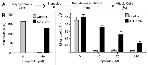
Figure 2 Inhibition of CHK1 by AZD7762 allows apoptosis. KG1a cells were treated with VP-16 (70 µM) for 1 h then released into fresh medium in the absence or presence of 300 nM AZD7762 for 20 h. Cells were then fixed and stained using cytotoxicity 3 HCS Reagent Kit, and the following indicators of cytotoxicity were measured by Cellomics Arrayscan instrument (Cellomics Inc.): mitochondrial membrane potential (A), mitochondrial cytochrome c release (B), membrane permeability (C). The arrayscan captures fluorescent images and performs automated image analysis using Compartmental analysis BioApplication. Statistical analyses were performed using a non-parametrical unpaired t-test (***p < 0.0001).
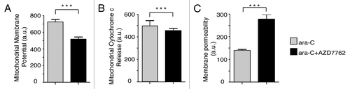
Figure 3 AZD7762 potentiated ara-C treatment on AML blast cells but not on normal granulomonocyte progenitors (CFU-GM). (A) AML blast cells (no. 1–11) and normal CD34+ HPC were grown in clonogenic assays in the presence of ara-C, alone or together with AZD7762 (10 nM). The clonogenic survival was assessed after 7 d and the IC50 for ara-C alone and in combination with AZD7762 was then calculated. The SF (sensitization factor) is the ratio between the IC50 for ara-C alone and in combination with AZD7762. (B) Correlation between SF and phospho-H2AX level (rMFI) (r = 0.85; ***p = 0.001). Statistical analyses were performed using a Pearson test.
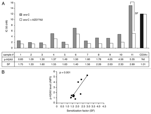
Figure 4 AZD7762 potentiated ara-C treatment on CD34+CD38−CD123+ stem cells compartments. (A) Primary leukemic cells from four patients were incubated for 48 h with or without 100 nM AZD7762 and/or 2 µM and then processed for apoptosis studies using annexin-V/7AAD staining after gating on CD34+CD38−CD123+. A representative AML sample is shown (B). Results are presented as percentage of survival cells.
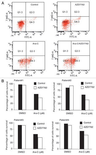
Table 1 Characteristics of AML patients