
Abstract
Targeting DNA repair with poly(ADP-ribose) polymerase (PARP) inhibitors has shown a broad range of anti-tumor activity in patients with advanced malignancies with and without BRCA deficiency. It remains unclear what role p53 plays in response to PARP inhibition in BRCA-proficient cancer cells treated with DNA damaging agents. Using gene expression microarray analysis, we find that DNA damage response (DDR) pathways elicited by veliparib (ABT-888), a PARP inhibitor, plus topotecan comprise the G1/S checkpoint, ATM, and p53 signaling pathways in p53-wildtype cancer cell lines and BRCA1, BRCA2 and ATR pathway in p53-mutant lines. In contrast, topotecan alone induces the G1/S checkpoint pathway in p53-wildtype lines and not in p53-mutant cells. These responses are coupled with G2/G1 checkpoint effectors p21CDKN1A upregulation, and Chk1 and Chk2 activation. The drug combination enhances G2 cell cycle arrest, apoptosis and a marked increase in cell death relative to topotecan alone in p53-wildtype and p53-mutant or -null cells. We also show that the checkpoint kinase inhibitor UCN-01 abolishes the G2 arrest induced by the veliparib and topotecan combination and further increases cell death in both p53-wildtype and -mutant cells. Collectively, PARP inhibition by veliparib enhances DDR and cell death in BRCA-proficient cancer cells in a p53-dependent and -independent fashion. Abrogating the cell-cycle arrest induced by PARP inhibition plus chemotherapeutics may be a strategy in the treatment of BRCA-proficient cancer.
Disclosure of Potential Conflicts of Interest
No potential conflicts of interest were disclosed.
Acknowledgments
The HCT116 p53-null isogenic cells were kindly provided by Dr. Bert Vogelstein at the Johns Hopkins Sydney Kimmel Comprehensive Cancer Center, Baltimore, MD. We thank Barbara Taylor at the Flow Cytometry Core, Center for Cancer Research, National Cancer Institute for her assistance in cell cycle analysis. This work was supported by the Division of Cancer Treatment and Diagnosis and the Center for Cancer Research, National Cancer Institute, National Institutes of Health.
Figures and Tables
Figure 1 Treatment effect on cell cycle and key cell cycle checkpoint proteins and kinases. (A) Cell cycle effects by treatments examined by flow cytometry. Cells were exposed to 1 µM veliparib, 10 nM topotecan alone or in combination for 24 h. Numbers above G1, S and G2 peaks indicate the percentage of cells in each phase of the cell cycle. Shown are the representative data of three independent experiments. (B) Protein gel blotting analyses of p21CDKN1A expression, activation of Chk1 and Chk2, and histone H3 phosphorylation (pH 3) after treatments for 24 h. Shown are the representative results of at least two independent experiments. ABT, veliparib; TPT, topotecan.

Figure 2 Enhancing effects of veliparib on apoptosis and cell death induced by topotecan. (A) Cleaved PARP detected by protein gel blotting 96 h after treatments. (B) Relative % changes in cell death in response to veliparib (gray bar), topotecan alone (white bar) or in combination (black bar) assessed by clonogenic assays. Shown are the representative data of two independent experiments. (C) Relationship between the cell death and G2 arrest (ratio of drug-treated/vehicle-treated) in response to veliparib, topotecan or in combination in HCT-116 p53+/+ (dot), HT-29 (diamond), MCF-7 (square), MDA-MB-231 (triangle), and p53−/− (star). ABT, veliparib; cl, cleaved; CC, correlation coefficient; TPT, topotecan.
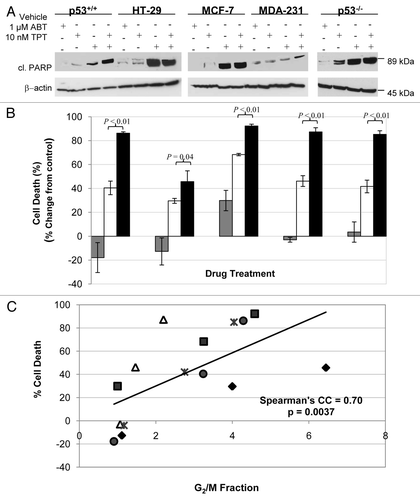
Figure 3 Effect of UCN-01 on the cell cycle distribution and cell death in cells treated with veliparib plus topotecan. After 24 h exposure to veliparib plus topotecan, the cultures were washed, and UCN-01 (100 nM) or vehicle was added for an additional 18 h. (A) Cell cycle effects of veliparib in combination with topotecan in the presence and absence of UCN-01. The numbers above G1 and G2 peaks indicate the percentage of cells in each phase of the cell cycle. (B) Percent change, relative to vehicle treatment without UCN-01, of cell death by veliparib plus topotecan without UCN-01 (gray bar), and with UCN-01(black bar) assessed by clonogenic assays. Shown are the representative data of two independent experiments. ABT, ABT-888; TPT, topotecan.
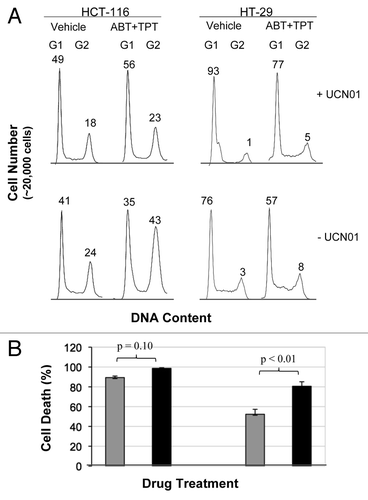
Table 1 The differentially expressed genes in the cell cycle
Table 2 The differentially expressed genes in role of BRCA1, BRCA2 and ATR in cancer susceptibility pathwayTable Footnote* by veliparib plus topotecan in p53-mutant lines