Abstract
Artemis, a member of the SNM1 gene family, is a multifunctional phospho-protein that has been shown to have important roles in V(D)J recombination, DNA double strand break repair, and stress-induced cell-cycle checkpoint regulation. We show here that Artemis interacts with the Cul4A-DDB1 E3 ubiquitin ligase via a direct interaction with the substrate-specificity receptor DDB2. Furthermore, Artemis also interacts with the CDK inhibitor and tumor suppressor p27, a substrate of the Cul4A-DDB1 ligase, and both DDB2 and Artemis are required for the degradation of p27 mediated by this complex. We also show that the regulation of p27 by Artemis and DDB2 is important for cell cycle progression in normally proliferating cells and in response to serum deprivation. These findings thus define a function for Artemis as an effector of Cullin-based E3 ligase-mediated ubiquitylation, demonstrate a novel pathway for the regulation of p27, and show that Cul4A-DDB1DDB2-Artemis regulates G1 phase cell cycle progression in mammalian cells.
Disclosure of Potential Conflicts of Interest
No potential conflicts of interest were disclosed.
Acknowledgments
This work was supported by National Cancer Institute grants CA096574 and CA097175.
Figures and Tables
Figure 1 Artemis interacts with DDB1 and DDB2 in vivo. (A) Gel stained with Coomassie blue showing proteins from a GST-Artemis pull-down experiment. GST-Artemis expressed in HEK293 cells was pulled-down by glutathione sepharose beads (left lane). A pull-down from untransfected cells was used as a control (middle lane). Labeled band indicate proteins identified by mass spectrometric analysis. “M” indicates marker proteins. (B) Immunoblot showing that Artemis co-IPs with DDB1 in the primary fetal lung cell line MRC5. (C) Immunoblot showing that endogenous DDB1 interacts with Artemis in vivo before and after UV or IR irradiation. HEK293 cells were treated with UV (25 J/m2), IR (10 Gy) or mock treated (UT). Cells were harvested for co-IP analysis two hours post-irradiation. “Pre” indicates pre-immune serum. (D) Reciprocal co-IP experiment showing that endogenous Artemis interacts with DDB1 in vivo before and after UV damage. HEK293 cells were treated with UV (25 J/m2) or left untreated. Cells were incubated for two hours after treatment and subsequently harvested for analysis. (E) Immunoblot showing that endogenous Artemis interacts with ectopically expressed DDB2. HEK293 cells were transfected with GST-DDB2, and 48 h later, cells were harvested for co-IP experiments. (F) Co-IP experiment showing the interaction between Artemis and DDB2. GsT-DDB2 and GFP-Artemis were co-transfected into HEK293 cells, and 48 h later, cells were harvested for analysis.
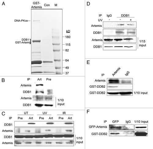
Figure 2 Artemis interacts directly with DDB2. (A and B) Artemis does not interact with DDB1 in vitro. Artemis and DDB1 labeled with 35S-methionine were expressed in vitro by transcription-coupled translation. Artemis and DDB1 were mixed and IPed with DDB1 antibody (A, upper part) or Artemis antiserum (B) as indicated. A control experiment shows the interaction between DDB1 and Cul4A (A, lower part). Bound proteins were eluted, separated by SDS-PAGE and detected by autoradiography. (C) Artemis directly interacts with GST-DDB2 in vitro. Artemis and GST-DDB2 were in vitro translated and labeled with 35S-methionine. Synthesized proteins were mixed and incubated as indicated for immunoprecipitation. Parts show the autoradiography of labeled proteins. (D) Purified recombinant Artemis and DDB2 directly interact. GST-Artemis, GST and MBP-DDB2 proteins were expressed and purified from E. coli. Purified proteins were mixed as indicated, and pull-down assays were performed and analyzed by immunoblotting.
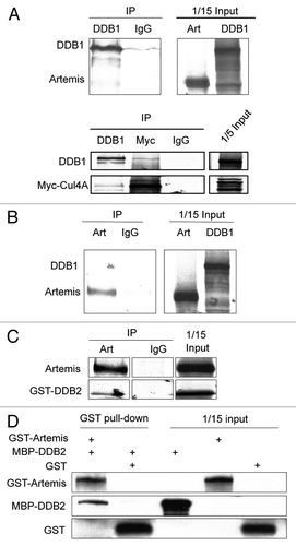
Figure 3 Artemis and DDB2 regulate p27 protein levels via a ubiquitin-mediated pathway. (A) P27 accumulates in Artemis or DDB2 depleted cells. HeLa cells were transfected with control (NS), Artemis or DDB2 siRNas. Forty-eight hours after transfection, cells were harvested and cell lysates were subjected to immunoblot analysis. Artemis-1 and Artemis-2, DDB2-1 and DDB2-2 indicate distinct siRNAs. As a control, 24 h after siRNA transfection cells were transfected with control plasmid DNA (C) or an Artemis construct refractory to Artemis siRNA (R). Cells were then incubated for an additional 24 h before harvesting. GAPDH and Actin indicate loading controls. “Q” indicates the relative band intensities of p27 normalized to GAPDH or actin levels. (B) p27 accumulates in Artemis null MEF cells. Lysates prepared from Artemis+/+ and Artemis−/− MEF cells were subjected to immunoblot analysis. (C) Overexpression of Artemis or DDB2 reduces p27 levels. HeLa cells were transfected with GST-Artemis (left part) or GST-DDB2 (right part) plasmid DNAs or treated with mock transfections. Forty-eight hours after transfection, cells were harvested and cell lysates were subjected to immunoblot analysis. (D) P27 mRNA is stable in Artemis or DDB2 depleted cells. mRNA was isolated from HeLa cells 48 h after transfection with control (NS), Artemis or DDB2 siRNAs. P27 mRNA levels were determined by real-time PCR. Results were normalized using GAPDH as an internal control. (E) Artemis and DDB2 regulate p27 protein levels through proteosome mediated degradation. HeLa cells were transfected with GST-Artemis or GST-DDB2 DNAs. After 48 h cells were treated with 20 mM MG-132, and 5 h later, cells were harvested and lysates subjected to immunoblot analysis. (F) Overexpression of either Artemis or DDB2 decreases the half-life of p27 compared with control. HeLa cells were transefected with GST-Artemis or GST control vectors. Thirty-six hours after transefection, cycloheximide (100 µg/ml) was added and the decrease in p27 protein level analyzed as a function of time by immunoblot analysis (left part). Quantitation of immunoblots are shown (right part). (G) Artemis and DDB2 promote ubiquitylation of p27 in vivo. HeLa cells were transfected with indicated amounts of Flag-p27, HA-Ub and GST-Artemis or GST-DDB2 DNAs. Cells were harvested 48 h after transfection, and an in vivo ubiquitination assay was performed using anti-Flag M2 agarose. Ubiquitylated proteins were detected using an HA antibody. Flag-p27 indicates a loading control at 10% of input. (H) Artemis promotes ubiquitylation of p27 independent of its phosphorylation on T187 or T157. HeLa cells were transfected with Flag-tagged wild-type (lane 1 and 4), T187A (lane 2) or T157A (lane3) p27, HA-Ub, GST-Artemis and GST-GUS. Forty-eight hours after transfection, an in vivo ubiquitylation assay was performed as indicated in .
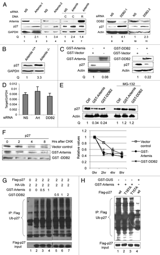
Figure 4 Artemis and DDB2 regulate p27 protein levels through the Cul4A-based E3 ligase complex. (A) Artemis and DDB2 are required for p27 degradation by the Cul4A-based E3 ligase complex. HeLa cells were transfected with control, Artemis or DDB2 siRNAs. After 24 h, cells were transfected with the indicated DNa, and 24 h later, cells were harvested for immunoblot analysis. (B) Cul4A is required for Artemis or DDB2-mediated p27 degradation. HeLa cells were transfected with control or Cul4A siRNAs, and 24 h later, cells were transfected with the indicated DNA. Cells were harvested after an additional 24 h for immunoblot analysis.
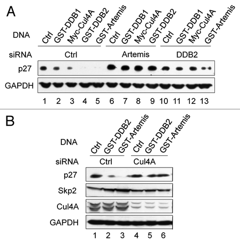
Figure 5 Artemis interacts with p27. (A) Artemis interacts with p27 in vivo. Immunoblot analysis showing co-IP experiment performed in lysates from HEK293 cells. (B) Artemis directly interacts with p27 in vitro. GST, GST-Artemis, MBP and MBP-p27 were expressed in E. coli. Purified proteins were mixed and subjected to GST pull-down assays. (C) Putative structural model for the roles of DDB2 and Artemis in the ubiquitylation of p27 by the Cul4A-DDB1 complex.
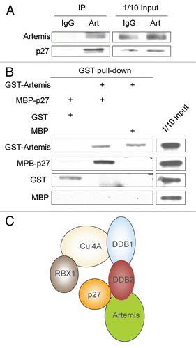
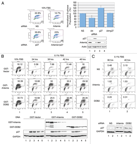
Figure 6 Artemis and DDB2 regulate cell cycle progression. (A) Depletion of Artemis causes a p27-dependent G1 arrest. hTERT-RPE1 cells were transfected with Artemis and/or p27 specific siRNA or non-specific (NS) siRNA. Forty-eight h after transfection, BrdU was added to label S-phase cells. Thirty minutes after incubation with BrdU, cells were harvested and fixed. Cell cycle analysis was performed by FACS (upper panel), and results from three independent experiments are shown graphically (lower left panel). Samples collected in the cell cycle analysis were subjected to immunoblot analysis for p27 and Artemis (lower right panel). Upper numbers in the panels indicate the percentage of S-phase cells. (B) Artemis and DDB2 affect the G1-G0 transition via regulation of p27. hTERT-RPE1 cells were transfected with GST-Artemis or GST-DDB2 constructs. Thirty h after transfection, cells were placed into DMEM F-12 media containing 0.1% FBs. Thirty minutes prior to harvest at the indicated times, BrdU was added to the media. Cell cycle profiles were determined by FACS analysis (upper panel). Lower panel: Samples collected in the cell cycle analysis were subjected to immunoblot analysis for p27 (lower panel). (C) Artemis and DDB2 affect the G1-G0 transition via regulation of p27. Upper panel: hTERT-RPE1 cells were transfected with Artemis, DDB2, or non-specific siRNAs (NS). Twenty-four h after transfection cells were placed into media containing 0.1% FBS for the indicated time. BrdU was added into the culture 30 min before harvest, and cell cycle analysis was performed by FACS (upper panel). Samples collected in the cell cycle analysis were subjected to immunoblot analysis for p27 (lower panel).