Abstract
Previous studies have demonstrated that curcumin induces mitochondria-mediated apoptosis. However, understanding of the molecular mechanisms underlying curcumin-induced cell death remains limited. In this study, we demonstrate that curcumin treatment of cancer cells caused dose- and time-dependent caspase-3 activation, which is required for apoptosis as confirmed using the pan caspase inhibitor, z-VAD. Knockdown experiments and knockout cells excluded a role of caspase-8 in curcumin-induced caspase-3 activation. In contrast, Apaf-1 deficiency or silencing inhibited the activity of caspase-3, pointing to a requisite role of Apaf-1 in curcumin-induced apoptotic cell death. Curcumin treatment led to Apaf-1 upregulation both at the protein and mRNA levels. Cytochrome c release from mitochondria to the cytosol in curcumin-treated cells was associated with upregulation of proapoptotic proteins such as Bax, Bak, Bid, and Bim. Crosslinking experiments demonstrated Bax oligomerization during curcumin-induced apoptosis, suggesting that induced expression of Bax, Bid, and Bim causes Bax-channel formation on the mitochondrial membrane. The release of cytochrome c was unaltered in p53-deficient cells, whereas absence of p21 blocked cytochrome c release, caspase activation, and apoptosis. Importantly, p21-deficiency resulted in reduced expression of Apaf-1 during curcumin treatment, indicating a requirement of p21 in Apaf-1 dependent caspase activation and apoptosis. Together, our findings demonstrate that Apaf-1, Bax, and p21 as novel potential targets for curcumin or curcumin-based anticancer agents.
Disclosure of Potential Conflicts of Interest
No potential conflicts of interest were disclosed.
Acknowledgments
We thank Drs. B. Vogelstein and Terry Beerman for providing reagents. This work was supported in part by a National Institutes of Health K01 Award CA123142 to D.C., NIH R01 award DK60632 and DK54909 to J.D.B. and National Cancer Institute Center Support Grant P30 CA016056 to the Roswell Park Cancer Institute. We apologize to those colleagues whose publications could not be cited due to space constraints.
Figures and Tables
Figure 1 Curcumin induces caspase 3 activation in multiple cell types. MDA-MB231 (A and B), PC3 (C and D) and LNCaP (E and F) were treated with curcumin (15 µM) for the indicated times. At the end of treatment, cells were harvested, washed with 1x PBS and lysed in caspase lysis buffer.Citation26 Equal amounts of protein were subjected to protein gel blotting for detection of caspase 3 processing or used for caspase 3 activity measurements (i.e., DEVDase activity). MDA, MDA-MB231 cells; and procasp-3, procaspase 3. Data are mean ± SD of three independent experiments. *p < 0.01.
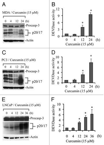
Figure 2 Curcumin induces caspase-dependent apoptosis. Cells were treated with curcumin (cur; 15 µM) or vehicle (control; DMSO) for 24 h, and percentage cell death was quantified using Trypan blue dye or cells were harvested and equal amounts of protein (50 µg) were subjected to caspase 3 activity (i.e., DEVDase activity) measurements. In some experiments, pan-caspase inhibitor (z-VAD; 50 µM) was added prior to curcumin treatment. Data are mean ± SD of three independent experiments. *p < 0.01.
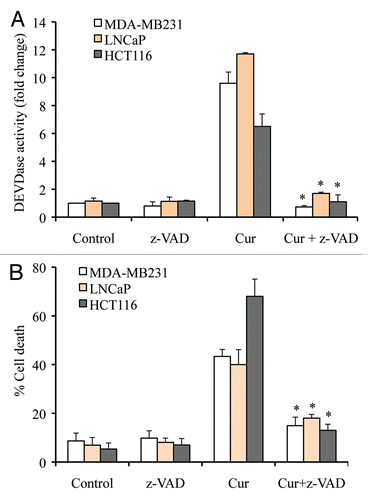
Figure 3 Caspase 8 deficiency does not modulate caspase activation and apoptosis. Jurkat WT and Jurkat caspase 8-/- cells (A and B) or Caspase 8-silenced LNCaP cells (C and D) were treated with curcumin (15 µM) for the indicated times. Percentage cell death was quantified using Trypan blue dye or cells were harvested and equal amounts of protein (50 µg) were subjected to caspase 3 activity measurements. Casp-8-/-, Jurkat caspase 8-/- cells; casp-8, procaspase 8. Data are mean ± SD of three independent experiments. *p < 0.01.
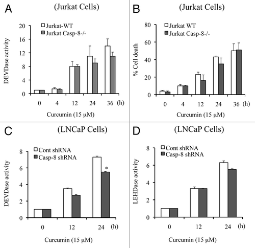
Figure 4 Apaf-1 deficiency inhibits curcumin-induced caspase 3 activation and apoptosis. MEFs WT and MEFs Apaf-1-/- cells (A and B) and Apaf-1 silenced LNCaP cells (C and D) were treated with curcumin (15 µM) for the indicated times. Percentage cell death was quantified using Trypan blue dye or cells were harvested and equal amounts of protein (50 µg) were subjected to caspase activity measurements. Data are mean ± SD of three independent experiments. *p < 0.01.
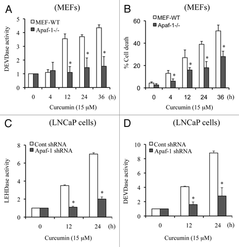
Figure 5 Curcumin treatment induces expression of Apaf-1 but downregulates FADD. MDA-MB231 cells were treated with curcumin (Cur) (15 µM) for the indicated times. At the end of treatment, samples were used for total RNA isolation or for the preparation of whole cell lysates. Equal amounts of protein were subjected to protein gel blotting for the detection of Apaf-1, FADD and Bak. A non-specific band serves as loading control. Isolated total RNAs were used to quantitate the expression of Apaf-1 and FADD using real-time PCR analysis. MDA, MDA-MB231 cells; NS, non-specific band serve as a loading control. Data are representative of three independent experiments.
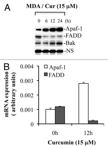
Figure 6 Curcumin induces cytochrome c release to the cytosol and is accompanied by nuclear fragmentation. (A) MDA-MB231 cells were treated with curcumin (15 µM) for indicated times. Cytosolic and mitochondrial fractions were isolated and equal amounts of protein were subjected to protein gel blotting for the detection of cytochrome c (Cyt. c), cytochrome c oxidase subunit II (COX II), heat shock protein 60 (Hsp60), actin or lactate dehydrogenase (LDH). Actin and Hsp60 serve as loading controls. (B) MDA-MB231 cells were treated with curcumin (15 µM) for 24 h. Following treatment, cells were incubated live with DAP I to label the nucleus and immunostained for cytochrome c (Cyt. c). Representative micrographs are shown; magnification bar represents 20 µM. Consistent with the protein gel analysis data, cytochrome c was released in individual cells as represented by diffuse cytochrome c staining. Apoptotic cells show fragmented or shiny nuclei with DAP I staining in (d). Data are representative of three independent experiments. Arrows indicate apoptotic cells showing cytochrome c release.
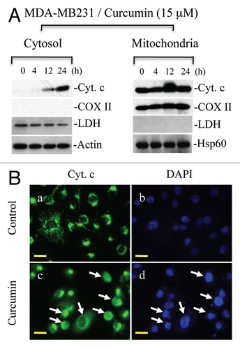
Figure 7 Curcumin induces upregulation of Bax, Bim and Bid, which trigger Bax oligomerization. (A) MDA-MB231 cells (MDA) were treated with curcumin (Cur) (15 µM) for the indicated times. Equal amounts of whole cell protein were separated on SDS-PA GE for detection of indicated proteins. Actin serves as loading control. (B) MDA-MB231 and HCT116 cells were treated with curcumin (15 µM) for the indicated times. In some treatment conditions, cells were pretreated with pancaspase inhibitor (z-VAD) one h prior to curcumin treatment. At the end of treatments, cells were cross-linked with BMH and subjected to protein gel blotting for the detection of Bax oligomers. Data are representative of three independent experiments.
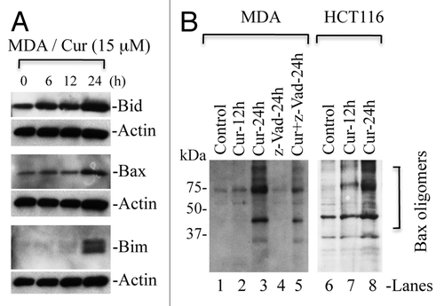
Figure 8 Curcumin induces Bax-dependent, p21-mediated cytochrome c release without p53 requirement. HCT116 WT (A), HCT116-p53-/- (B), HCT116-Bax-/- (C) and HCT116-p21-/- (D) cells were treated with curcumin (15 µM) for the indicated times. Cytosolic and mitochondrial fractions were isolated and equal amounts of protein were subjected to protein gel blotting for the detection of cytochrome c (Cyt. C), cytochrome c oxidase subunit II (COX II), heat shock protein 60 (Hsp60) and actin. Actin and Hsp60 serve as loading controls. *represents a non-specific band.
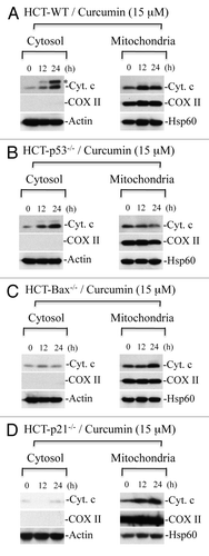
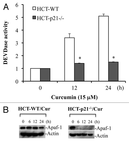
Figure 9 p21 deficiency reduces Apaf-1 expression and inhibits curcumin-induced caspase activation. HCT116 WT and HCT116-p21-/- cells were treated with curcumin (Cur) (15 µM) for the indicated times. At the end of treatment, cells were harvested and equal amounts of protein (50 µg) were subjected to caspase 3 activity measurements, or protein gel blotting for Apaf-1. Actin serves as a loading control. HCT-WT, HCT116-WT cells; HCT-p21-/-, HCT116-p21-/--deficient cells. Data are mean ± SD of three independent experiments. *p < 0.01.