Abstract
Tight regulation of the cell cycle and DNA repair machinery is essential for maintaining genome stability. The APC/CCdh1 ubiquitin ligase complex is a key regulator of protein stability during the G1 phase of the cell cycle. APC/CCdh1 regulates and promotes the degradation of proteins involved in both cell cycle regulation and DNA repair. In a recent study, we identified a novel APC/CCdh1 substrate, the ubiquitin protease USP1. USP1 is a critical regulator of both the Fanconi anemia (FA) and translesion synthesis (TLS) DNA repair pathways. Here, we provide additional mechanistic insights into the regulation of USP1 during the cell cycle. Specifically, we demonstrate that USP1 is phosphorylated in mitosis by cyclin-dependent kinases (Cdks), and that this phosphorylation event may prevent premature degradation of USP1 during normal cell cycle progression. Finally, we provide a unifying hypothesis integrating the role of G1-specific proteolysis of USP1 with the regulation of the transcriptional repressors, Inhibitor of DNA-binding (ID) proteins.
Acknowledgments
We especially would like to thank Luca Colnaghi and Miklos Bekes for helpful discussions of the manuscript. We also thank members of the T. Huang, M. Pagano, D. Bar-Sagi and D. Reinberg laboratories for their valuable tools, reagents, equipment and technical assistance. This work was supported by a National Institute of Health grant to T.T. Huang (R01-GM084244).
Figures and Tables
Figure 1 Multiple mechanisms regulate USP1 activity. (A) USP1 requires the association of UAF1 for its full enzymatic activity and protein stability. (B) Upon UV DNA damage, USP1 is auto-cleaved and degraded by the proteasome by an unknown ubiquitin E3 ligase. (C) APC/CCdh1 binds to and degrades USP1 during the G1 phase of the cell cycle.
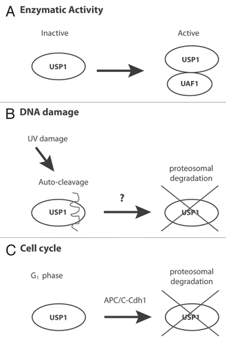
Figure 2 Inhibition of Emi1 or Cdks can destabilize USP1. (A) U2OS cells were transfected for 48 h with a control (Ctrl) siRNA oligo (Qiagen AllStar Neg siRNA) or a siRNA oligo directed against Emi1 (5′-CAT GTT CAT TCC GGA CTT AAA-3′). Samples were collected, lysed and analyzed by protein gel blot with the indicated antibodies: Plk1 (Abcam), Cdc6 (Santa Cruz), Emi1 (Invitrogen), Cdk2 (Bethyl), Cdh1 (CalBiochem), Tubulin (Abcam). (B) U2OS cells were left untreated (UN) or treated with Roscovitine (CalBiochem) (5 uM) for the indicated time points, analyzed by protein gel blot and probed with the indicated antibodies. (C) U2OS cells were transfected with the indicated siRNAs as in (A) or Cdh1 (AAT GAG AA G TCT CCC AGT CAG) and treated for 6 h with Roscovitine (5 uM). Samples were analyzed by protein gel blot and probe with indicated antibodies.
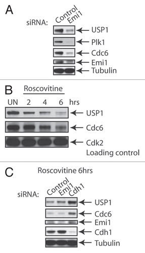
Figure 3 Cdk-dependent phosphorylation of USP1 on S313. (A) Schematic diagram of USP1 catalytic domains, Cdh1 recognition region and putative Cdk phosphorylation site S313 on human USP1. (B) U2OS cells were transfected with Myc-USP1 WT or Myc-USP1 S313A using Fugene6 transfection reagent (Roche Applied Science). Samples were lysed and immunoprecipitated with anti-Myc antibody (Santa Cruz) and protein gel blot was performed using anti-Myc and P-S313 USP1 antibodies (Thermo Scientific). Lysing, immunoprecipitation and washing buffers were done using the low IPB buffer [25 mM Tris, pH 7.5, 150 mM NaCl, 2 mM EDTA and 0.5% NP-40 with protease inhibitor cocktail (Roche)]. (C) U2OS cells were synchronized in M phase by incubating cells with Nocodazole (0.1 µg/ml) for 12–16 h. After 12-16 h, mitotic cells were shaken off and collected and washed twice with 1x PBS, then plated with fresh media and collected for the indicated time points. Samples were analyzed by protein gel blot and probe with the indicated antibodies: P-S313 USP1 antibody (Thermo Scientific), H3 Ser10 (Millipore) and others described above. (D) U2OS cells synchronized in M phase, as in (C), and were treated (or left untreated) with Cdk1 inhibitor RO-3306 (Calbiochem) (10 uM) in the presence or absence of MG132 (10 uM). Samples were analyzed by protein gel blot using the indicated antibodies. (E) U2OS cells were transfected as in (C), with Myc-USP1 WT, S313A or S313E, then cells were split, left unsynchronized (AS) or synchronized in mitosis (M). Samples were analyzed by protein gel blot and probe with the indicated antibodies.
![Figure 3 Cdk-dependent phosphorylation of USP1 on S313. (A) Schematic diagram of USP1 catalytic domains, Cdh1 recognition region and putative Cdk phosphorylation site S313 on human USP1. (B) U2OS cells were transfected with Myc-USP1 WT or Myc-USP1 S313A using Fugene6 transfection reagent (Roche Applied Science). Samples were lysed and immunoprecipitated with anti-Myc antibody (Santa Cruz) and protein gel blot was performed using anti-Myc and P-S313 USP1 antibodies (Thermo Scientific). Lysing, immunoprecipitation and washing buffers were done using the low IPB buffer [25 mM Tris, pH 7.5, 150 mM NaCl, 2 mM EDTA and 0.5% NP-40 with protease inhibitor cocktail (Roche)]. (C) U2OS cells were synchronized in M phase by incubating cells with Nocodazole (0.1 µg/ml) for 12–16 h. After 12-16 h, mitotic cells were shaken off and collected and washed twice with 1x PBS, then plated with fresh media and collected for the indicated time points. Samples were analyzed by protein gel blot and probe with the indicated antibodies: P-S313 USP1 antibody (Thermo Scientific), H3 Ser10 (Millipore) and others described above. (D) U2OS cells synchronized in M phase, as in (C), and were treated (or left untreated) with Cdk1 inhibitor RO-3306 (Calbiochem) (10 uM) in the presence or absence of MG132 (10 uM). Samples were analyzed by protein gel blot using the indicated antibodies. (E) U2OS cells were transfected as in (C), with Myc-USP1 WT, S313A or S313E, then cells were split, left unsynchronized (AS) or synchronized in mitosis (M). Samples were analyzed by protein gel blot and probe with the indicated antibodies.](/cms/asset/d1ff67f0-f5fe-4db9-b10f-c456a413726f/kccy_a_10918501_f0003.gif)
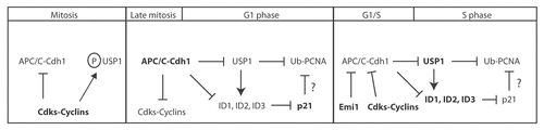
Figure 4 Model showing how USP1-mediated events are regulated during normal cell cycle progression. Schematic diagram of the proposed model of how different cell cycle stages can affect USP1 protein stability, regulation of ID proteins and PCNA-directed DNA repair. Briefly, in M phase, both USP1 and APC/CCdh1 are phosphorylated by Cdks, which prevents USP1 from being prematurely targeted for degradation by the APC/CCdh1. In late M and early G1, USP1 and APC/CCdh1 become dephosphorylated, which leads to the degradation of both USP1 and cyclins. USP1 normally protects ID proteins from ubiquitin-mediated degradation. However, without USP1, ID proteins become subsequently degraded. Loss of ID proteins prevents transcriptional repression of p21, leading to p21 protein accumulation and possible inhibition of TLS activity on PCNA. During the G1-S or S-phase entry, levels of cyclins rise to inhibit APC/CCdh1, which lead to the accumulation of USP1 and presumed stabilization of ID proteins.