Abstract
Disruption of chromatin organization during replication poses a major challenge to the maintenance and integrity of genome organization. It creates the need to accurately reconstruct the chromatin landscape following DNA duplication but there is little mechanistic understanding of how chromatin based modifications are restored on newly synthesized DNA. ATP-dependent chromatin remodeling activities serve multiple roles during replication and recent work underscores their requirement in the maintenance of proper chromatin organization. A new component of chromatin replication, the SWI/SNF-like chromatin remodeler SMARCAD1, acts at replication sites to facilitate deacetylation of newly assembled histones. Deacetylation is a pre-requisite for the restoration of epigenetic signatures in heterochromatin regions following replication. In this way, SMARCAD1, in concert with histone modifying activities and transcriptional repressors, reinforces epigenetic instructions to ensure that silenced loci are correctly perpetuated in each replication cycle. The emerging concept is that remodeling of nucleosomes is an early event imperative to promote the re-establishment of histone modifications following DNA replication.
Acknowledgments
We are grateful to Florence Cammas, Inserm, France, for providing F9 cells that express low levels of KAP1/TIF1β (TIF1β-/-/rTA-f.TIF1β). We thank Wendy Dean, Colin Dingwall, Louise Matheson, Fatima Santos and Nicola Stead for comments on the manuscript. This work was supported by the UK Biotechnology and Biological Sciences Research Council (BBSRC), the UK Medical Research Council, the European Union FP6 Network of Excellence “The Epigenome” and a BBSRC studentship to S.R.
Figures and Tables
Figure 1 ATP-dependent chromatin remodeling complexes play important roles during all steps of replication: they facilitate the disassembly of nucleosomes ahead of the replication fork, efficient progression of replication, subsequent proper assembly of chromatin onto newly synthesized DNA, the copying of epigenetic information onto the replicated chromatin (PTM: post-translational modification) as well as the repair of DNA during replication.
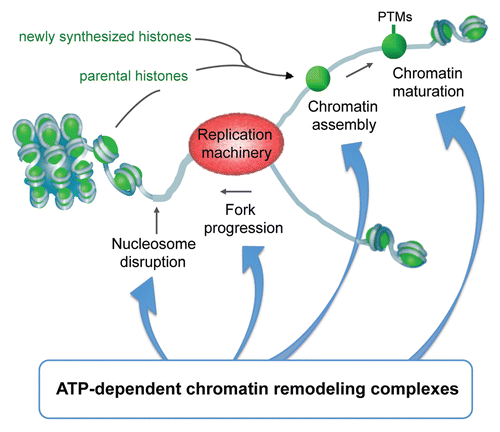
Figure 2 SMARCAD1 knockdown (KD) affects histone modifications and protein occupancy at pericentric repeats. Chromatin immunoprecipitation of H3ac, H3K9me3, HDAC1 and KAP1 at satellite repeats from SMARCAD1 KD and control HeLa cells. %IP from KD cells is shown relative to %IP from control cells which is normalized to 100. Error bars denote standard deviation from 3 independent experiments. Primers: Sat1 (this study) forward 5′-TTG AAG GTA TAT TCA TAC TGG CC-3′ reverse 5′-TTC AAA GGT ACT CTG CTT GGT ACA-3′ NBL2Citation30 forward 5′-TCC CAC AGC AGT TGG TGT TA-3′ reverse 5′-TTG GCA GAA ACC TCT TTG CT-3′.
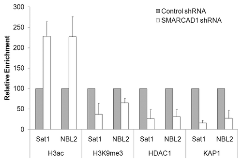
Figure 3 Summary of SMARCAD1 interacting proteins (listed in full in ref. Citation9). (A) Several SMARCAD1 interaction partners have overlapping functions in gene silencing and heterochromatin formation, replication and repair. (B) A number of chromatin remodelers co-purify with SMARCAD1.
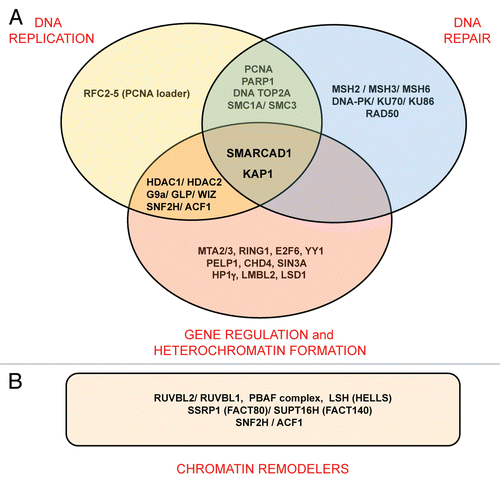
Figure 4 Model of SMARCAD1 function in chromatin replication: SMARCAD1 is recruited to replication sites by PCNA where it functions in a complex with KAP1, HDAC1, HDAC2 and the histone methyltransferase G9a/GLP. Deacetylation of newly assembled histones is facilitated by SMARCAD1 nucleosome remodeling and primes new nucleosomes for further modifications, promoting the inheritance of H3K9 methylation and the formation of heterochromatin.
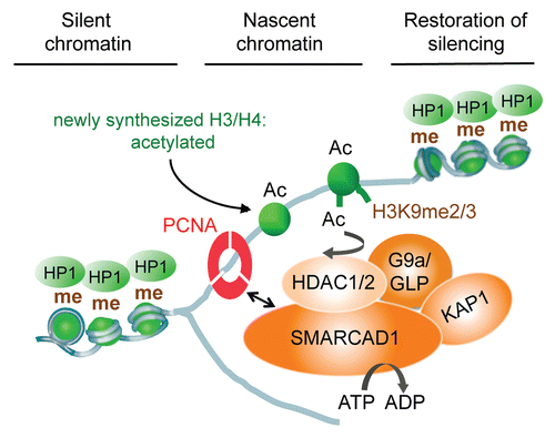
Figure 5 Multiple independent regions of SMARCAD1 co-localize with PCNA. (A) Cartoon representation of tagged SMARCAD1 protein and truncations, amino acids (aa) are indicated. (B) Confocal microscopy of SMARCAD1 knockdown HeLa cells expressing Venus-SMARCAD1 proteins and CFP-PCNA. Panels on the right shows a merge of the PCNA and SMARCAD1 channels. Images depict the autofluorescence of the transfected proteins in fixed cells. Representative cells are shown, images were pseudo-colored and adjusted for brightness and contrast.
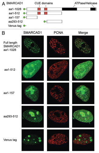
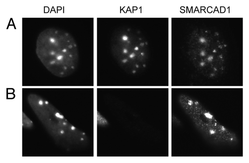
Figure 6 Localization of SMARCAD1 to pericentric heterochromatin is not dependent on KAP1 levels. (A) F9 embryonic carcinoma cells and (B) F9 cells that were engineered to express low levels of KAP1/TIF1β (TIF1β-/-/rTA-f.TIF1β) were differentiated for 7 d by exposure to 1 µM retinoic acid as described by Cammas et al. representative cells stained for KAP1 (ab22553) and SMARCAD1 Citation9 are shown, images were adjusted for brightness and contrast. DAPI bright foci mark pericentric heterochromatin.