
Abstract
Radiotherapy is the primary treatment for nasopharyngeal carcinoma (NPC), but radioresistance severely reduces NPC radiocurability. Here, we have established a radio-resistant NPC cell line, CNE-2R, and investigate the role of miRNAs in radioresistance. The miRNAs microarray assay reveals that miRNAs are differentially expressed between CNE-2R and its parental cell line CNE-2. We find that miR-205 is elevated in CNE-2R. A target prediction algorithm suggests that miR‑205 regulates expression of PTE N, a tumor-suppressor. Introducing miR-205 into CNE-2 cells suppresses PTE N protein expression, followed by activation of AKT, increased number of foci formation and reduction of cell apoptosis postirradiation. On the other hand, knocking down miR-205 in CNE-2R cells compromises the inhibition of PTE N and increases cell apoptosis. Significantly, immunohistochemistry studies demonstrate that PTE N is downregulated at late stages of NPC, and that miR-205 is significantly elevated followed the radiotherapy. Our data conclude that miR-205 contributes to radioresistance of NPC by directly targeting PTE N. Both miR-205 and PTE N are potential predictive biomarkers for radiosensitivity of NPC and may serve as targets for achieve successful radiotherapy in NPC.
Disclosure of Potential Conflicts of Interest
No potential conflicts of interest were disclosed.
Acknowledgements
This work was supported in part by the NIHRO1CA (089266), Cancer Center Core Grant (CA16672), National Natural Science Foundation of China (NO: 30670627; 30870745; 81071837), National Natural Science Foundation of Guangdong Province, China (NO: 9251008901000005; 06021210), the International Program Fund of 985 Project, Sun Yat-Sen University, China and Exchange Visitor Program (NO: P-1-10254), the University of Texas MD Anderson Cancer Center.
Figures and Tables
Figure 1 CNE-2R is radio-resistant. (A) CNE-2R is more IR resistant than CNE-2. Indicated cell lines were treated with indicated amounts of irraditiation and forci-formation was indicated. (B) CNE-2R has reduced numbers of forci formation. Indicated cells were plated in triplicate and exposed to a range of IR doses (0–6 Gy). The numbers of foci-formation were presented as bar graphs. (C) CNE-2R has reduced survival fraction. Survival fractions were calculated by dividing the number of colonies formed after IR by the corresponding number of colonies formed without IR from experiments in (B). (D) Growth of CNE-2R is not affected by IR. CNE-2 and CNE-2R plated in 24-well culture plates were exposed to IR with 2 Gy and cell growth was monitored by counting cell numbers. (E) CNE-2R is resistant to IR-induced cell death. The cells were treated with or without 10 Gy IR for analysis of apoptosis of CNE-2 and CNE-2R cells after IR. At 48 h post-irradiation, the cells were stained with PI and the percentage of sub-G1 cells was measured by flow cytometry in three independent experiments. ns, no significance; *p < 0.05; ***p < 0.001, ****p < 0.0001. Data were presented as mean ± SD.
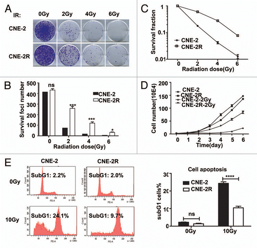
Figure 2 miR-205 is elevated in radio-resistant NPC cells. (A) miR expressing signals from CNE-2 or CNE-2R. miRNAs from CNE-2 or CNE-2R were labeled with Cy3 or Cy5 respectively. (B) Scatter plot of miR expression profiles in radiosensitive CNE-2 (x-axis) and radio-resistant CNE-2R cells (y-axis). The miRs differentially expressed with statistical significance were marked in red. (C) Differential miR expression between CNE-2 and CNE-2R. Relative expression levels of several representative miRs with differential expression levels were presented. (D) Relative miR-205 expression level is different between CNE-2 and CNE-2R after IR. Quantification by qRT-PCR of miR-205 expression level in CNE-2R and CNE-2 cells before and after IR. U6 was used for normalization. Data were presented as mean ± SEM (n = 3). ns, no significance; *p < 0.05; **p < 0.01; ***p < 0.001.
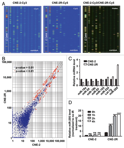
Figure 3 Expression of miR-205 in radio-sensitive cells leads to IR resistance. (A) miR-205 overexpression in CNE-2 cells. CNE-2 cells were transfected with miR-205 expressing vector. Expression of miR-205 was quantitated by q-RTPCR. Expression of miR-205 in the transduced cells was assessed by qRT-PCR with U6 RNA as an internal control. (B) CNE-2 overexpressing miR-205 becomes more IR resistant. Indicated miR-205-overexpressing CNE-2 cells were plated in triplicate and exposed to a range of IR doses (0–6 Gy). The forci-formation was indicated. (C) miR-205-overexpressing CNE-2 cells have induced numbers of forci formation. The numbers of foci-formation were presented as bar graphs. (D) miR-205-overexpressing CNE-2 cells have increased survival fraction. Survival fractions were calculated as described above based on the data from experiments in (C). (E) miR-205-overexpressing CNE-2 cells is resistant to IR-induced cell death. Indicated cells were treated with or without 10 Gy IR and the cells were stained with PI and the percentage of sub-G1 cells was measured by flow cytometry in three independent experiments. The percentage of sub G1 cells was presented as bar graphs.
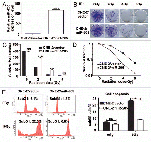
Figure 4 Suppression of Mir 205 in CNE-2R cells leads to radiosensitivity to IR-regulated cell death. (A) Lentiviral anti-miR-205 leads to miR-205 suppression in CNE-2R cells. CNE-2R cells were transfected with lenti-virus based copGFP-anti-miR-205 vector. Expression of miR-205 was quantitated by q-RTPCR. (B) CNE-2R overexpressing anti-miR-205 becomes more IR sensitive. Indicated anti-miR-205-overexpressing CNE-2R cells were plated in triplicate and exposed to a range of IR doses (0–6 Gy). The forci-formation was indicated. (C) anti-miR-205-overexpressing CNE-2R cells have reduced numbers of forci formation. The numbers of foci-formation were presented as bar graphs. (D) anti-miR-205-overexpressing CNE-2R cells have reduced survival fraction. Survival fractions were calculated as described above based on the data from experiments in (C). (E) anti-miR-205-overexpressing CNE-2R cells is sensitive to IR-induced cell death. Indicated cells were treated with or without 10 Gy IR and the cells were stained with PI for measuring the percentage of sub G1 cells. Three independent experiments were done. The percentage of sub G1 cells in experiment was presented as bar graphs. ns, no significance; ****p < 0.0001. (F) anti-miR-205-overexpressing CNE-1 cells is sensitive to IR-induced cell death. Cells were treated with or without 10 Gy IR and the cells were stained with annexin V for measuring the percentage of apoptotic cells. The percentage of apoptotic cells in experiment was presented as bar graphs. ns, no significance; ****p < 0.0001.
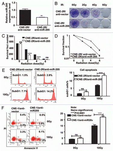
Figure 5 Suppressing the expression of miR-205 compromises the IR-induced downregulation of PTEN. (A) MiR-205 represses PTEN expression by targeting the 3′-UTR of PTEN mRNA. An miR-205 binding site within PTEN 3′UTR that is conserved in different species was predicted by TargetScan. The “seed” sequences of miR-205 complementary to PTEN are shown in red box. (B) Hybridization of miR-205 (green) and PTEN mRNA (red) was predicted by RNAhybrid software. The minimum free energy (mfe) required for this hybridization is indicated. (C) Expression of Mir 205 in CNE-2 cells leads to downregulation of PTEN and increased akt activity. Levels of PTEN protein expression and phosphorylation of Akt after transfection of miR-205 into CNE-2 cells were demonstrated by indicated antibodies. (D) PTEN mRNA levels is downregulated after miR-205 transfection. Gene expression was measured by qRT-PCR. Data were presented as mean ± SEM (n = 3). ****p < 0.0001. (E) Reporter gene assay. pMIR-REPORT, 3′-PTEN-miR-205 and the mutant reporter 3′-PTEN-miR-205-deletion were transfected into CNE-2/vector and CNE-2/miR-205 cells. Luciferase reporter gene activities were assayed. Data were presented as mean ± SEM of 3 experiments. ***p < 0.001. (F) Suppressing the expression of Mir-205 compromises the IR-induced downregulation of PTEN. Indicated cells were irradiated with 10 Gy IR or without. The lysates were immunoblotted with indicated antibodies.
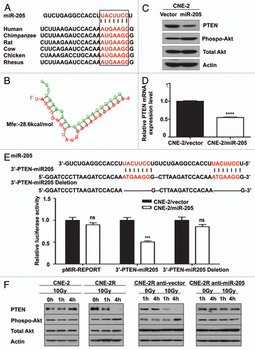
Figure 6 Inhibition of PI3K/Akt signaling pathway can reverse radioresistance of NPC. Indicated cells were treated with or without 10 µM Ly294002 in the presence of IR. The cells were stained with annexin V for measuring the percentage of apoptotic cells. The percentage of apoptotic cells in experiment was presented as bar graphs. ns, no significance; ****p < 0.0001.
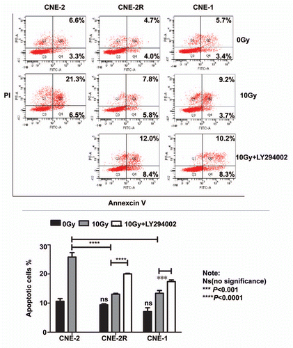
Figure 7 PTEN is downregulated in late stages of NPC. (A) High PTEN expression correlates with late stages in NPC. NPC cancer tissues were immunostained with anti-PTEN. Micrographs of two representative NPC cancer specimens were shown (original magnification, 200x). (B) Upregulaion of miR-205 is observed in NPC tissue after radiotherapy. qRT-PCR was used to quantify miR-205 expression levels in the NPC tissues obtained before and after radiotherapy. Data were presented as mean ± SEM of 3 experiments. ***p < 0.001, compared with before radiotherapy.
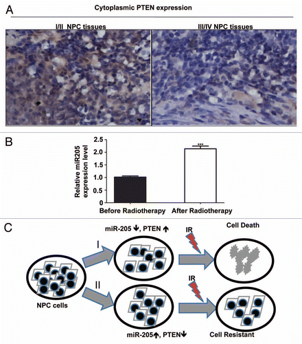
Table 1 The difference of PTEN expression in NPC tissues with different clinical stages