Abstract
Many cancer patients are treated with a combination of anticancer drugs. Here, we discuss the importance of drug scheduling and the need for studies that investigate the optimal timing of the various anticancer drugs. Positron emission tomography (PET) using radiolabeled anticancer drugs could be an important tool for those studies.
Introduction
Over the past decades, the development of new drugs and new treatment modalities has improved the prospect of cancer patients. To date, numerous drugs have been introduced. This number is still increasing, and an ever increasing number of drugs will become available for the treatment of cancer. A new anticancer drug is often added to an existing treatment strategy, as combination therapies reduce the development of drug resistance, leading to synergyCitation1,Citation2 and a lower rate of treatment failure. As a result, an increasing number of combination therapies are under investigation for clinical implementation. Usually, the feasibility of new combination strategies is evaluated in phase-I, phase-II and eventually in phase-III trials. The purpose of phase-I studies is to assess whether patients can tolerate a new combination of anticancer drugs, and therefore both incidence and severity of experienced adverse events are scored. In addition, pharmacokinetic analyses are performed to determine the effects of combination strategies on plasma levels of the drugs and their metabolites. In a phase-II trial, the new combination can be investigated in specific cancer types. Furthermore, it is evaluated whether it is feasible to test the new combination in a larger phase-III trial. When the new combination successfully passes phase-I and phase-II trials, it finally reaches a phase-III trial. In these phase-III trials, the additional value of the new drug is compared with standard therapy. In a phase-III trial, patients are randomized to a treatment with or without the new drug, and the clinical outcomes of the two arms, usually defined as overall survival, are compared. Based on positive results of a randomized phase-III trial, the new drug can be registered and find its way to the clinic. Within the context of a new combination, however, optimal scheduling of drugs often is not investigated. In this perspective, we will discuss whether scheduling of anticancer drugs may affect efficacy of anticancer treatment. In addition, we will illustrate how positron emission tomography (PET) using radiolabeled anticancer drugs may be an important method to elucidate the importance of drug scheduling.
Anti-Angiogenic Drugs for Treatment of Cancer
Among approved anticancer agents, drugs that target tumor angiogenesis are currently widely prescribed for the treatment of several advanced malignancies.Citation3-Citation9 Angiogenesis is essential for survival of malignant tumorsCitation10,Citation11 and has become an important target in the treatment of cancer. In particular, drugs have been developed that target pathways of the vascular endothelial growth factor (VEGF) and its receptors (VEGFR).Citation12 VEGF is overexpressed in malignant tumors, and it is an important growth factor for tumor angiogenesis.Citation13 As single-agents, however, anti-angiogenic drugs usually are not sufficiently effective for the treatment of most malignancies. Consequently, anti-angiogenic drugs often are combined with conventional chemotherapy, as this strategy has shown additional value in several cancers.Citation3,Citation4,Citation7 However, the story of the anti-angiogenic drug bevacizumab has shown that the place of anti-angiogenic drugs in the clinic is not certain, as an initially obtained approval can, in case of disappointing results, be revoked by the Food and Drug Administration (FDA). In 2008, bevacizumab, a humanized monoclonal antibody that targets circulating VEGF,Citation14 was granted accelerated approval for first-line treatment of human epidermal growth factor receptor 2 (HER2)-negative metastatic breast cancer.Citation5,Citation15 At the time, addition of bevacizumab to paclitaxel was approved on the basis of a significant improvement in progression-free survival as compared with paclitaxel alone.Citation5,Citation15 Later studies, however, could not confirm the increase in progression-free survival, which is a surrogate clinical end point, and did not show evidence for improved overall survival.Citation16 As a result, the FDA concluded that bevacizumab was not shown to be effective in patients with (HER2)-negative metastatic breast cancer, and its approval for this patient population was revoked.Citation16 It has been suggested that the discrepant results for bevacizumab in the treatment of metastatic breast cancer may be explained by the different chemotherapy regimens used and that the additional value of bevacizumab may be chemotherapy-specific.Citation17 More importantly, scheduling of anticancer drugs may also affect the delivery of chemotherapy to tumors, potentially leading to differences in efficacy of combination therapy. In the latter case, PET using radiolabeled anticancer drugs can be used to investigate this potential mechanism.
PET Using Radiolabeled Anticancer Drugs
In oncology, PET scans using fluorine-18-labeled deoxy-glucose [2-(18F)fluoro-2-deoxy-D-glucose or (18F)FDG] are used routinely for diagnosis and staging of numerous malignancies.Citation18 In addition, this non-invasive imaging technique can also be applied to monitor pharmacokinetics and pharmacodynamics of anticancer drugs in vivo.Citation19 To this end, a tracer dose (microdose) of an anticancer drug is radiolabeled with a short-lived positron emitting radionuclide such as carbon-11 or fluorine-18. After intravenous administration of a radiolabeled anticancer drug, its uptake in tumor tissue can be measured non-invasively using PET. Preliminary PET studies using fluorine-18-labeled 5-fluorouracil [(18F)5-FU],Citation20,Citation21 fluorine-18-labeled tamoxifen [(18F)fluorotamoxifen]Citation22 and carbon-11-labeled docetaxel [(11C)docetaxel]Citation23 have demonstrated that high tumor uptake of the radiolabeled anticancer drug was associated with improved tumor response. These studies indicate that radiolabeled anticancer drugs may be useful for predicting outcome prior to start of treatment. In addition, such PET scans can be useful to investigate whether tumor uptake of a radiolabeled drug is affected by administration of another drug.
Role of PET for Evaluation of Drug Scheduling
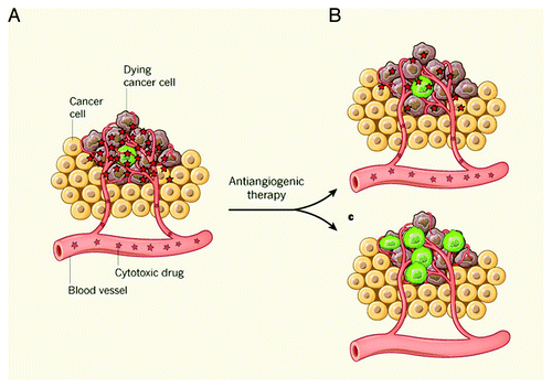
The effects of drug scheduling can be explored using PET and radiolabeled anticancer drugs. On one hand, PET using radiolabeled drugs can contribute to conventional pharmacokinetic studies by monitoring metabolism of the radiolabeled drug. For example, administration of eniluracil, an inhibitor that suppresses catabolism of 5-FU by inactivating dihydrouracil dehydrogenase in the liver, decreased (18F)5-FU uptake in normal liver and kidneys, increased plasma levels of uracil and non-metabolized (18F)5-FU and improved uptake of radioactivity in tumors, reflecting enhanced tumor uptake of (18F)5-FU and/or its radiolabeled anabolites.Citation24-Citation27 On the other hand, effects of other (anticancer) drugs on the delivery of radiolabeled drugs to tumors can be evaluated. Several PET studies have applied this concept to (18F)5-FU.Citation28,Citation29 It has been shown that interferon-α increases perfusion and uptake of (18F)5-FU in several malignant tumors, whereas N-phosphonacetyl-L-aspartate (PALA) induces a decrease in these parameters.Citation29 As drug delivery to tumors may be related to tumor perfusion,Citation23 administration of drugs that affect tumor perfusion is of interest.Citation30,Citation31 Tumor perfusion can be assessed using PET and radioactive water [(15O)H2O], which has a half-life of ~2 min, enabling sequential PET scans using both (15O)H2O and a radiolabeled anticancer drug.Citation32 For example, (15O)H2O PET showed that administration of angiotensin II, a drug that causes hypertension, selectively increases perfusion in hepatic tumors.Citation33-Citation35 As mentioned above, effects of anti-angiogenic drugs on drug delivery to tumors is of special interest. Recently, we have investigated the effect of bevacizumab on delivery of (11C)docetaxel to tumors.Citation36 Non-small cell lung cancer patients underwent PET scans with (15O)H2O and (11C)docetaxel prior to and at 5 h and 4 d after infusion of bevacizumab. Within 5 h, both perfusion and (11C)docetaxel uptake in tumors had significantly decreased, and these effects persisted after 4 d. Reduction in (11C)docetaxel delivery to tumors was accompanied by rapid reduction in circulating levels of VEGF, but was not associated with changes in cardiovascular parameters, including blood pressure, cardiac output and capillary density in the skin. According to these results, a 20% decline in (18F)5-FU uptake was also measured in human colorectal cancer at 24 h after administration of bevacizumab.Citation37 Besides a decrease in drug delivery (), the decreased tumor perfusion results in oxygen deficiency (also called hypoxia) in tumors, which, in turn, may result in more aggressive cancer cell populations that have an increased capacity to spread to other organs ().Citation38,Citation39 Although the mentioned PET studies do not prove that the bevacizumab-induced reduction in delivery of chemotherapy affects efficacy of the drugs, they highlight the importance of drug schedulingCitation40 and show that the administration of anti-angiogenic drugs may be considered after the cytotoxic drugs, as the immediate decrease in tumor perfusion may decrease clearance of cytotoxic drugs from tumor tissue. In addition, these studies show the promising application of radiolabeled anticancer drugs for characterizing the effects of drug scheduling on their delivery to tumors, in particular, that of anti-angiogenic drugs. Apart from the applications mentioned above, effects of inhibitors of efflux transporters, such as ABCB1, are also of interest. Overexpression of ABCB1 is found in many drug-resistant tumorsCitation41 and may decrease drug uptake in tumors. Consequently, inhibitors of ABCB1 may improve drug uptake in tumors. In addition, inhibitors of ABCB1 may decrease the rapid clearance of drugs from blood,Citation42,Citation43 as ABCB1 is also extensively expressed in intestine and the biliary system,Citation44 and may contribute to drug elimination.Citation45 Therefore, PET studies using both radiolabeled anticancer drugs and tracers that can show in vivo functionality of multidrug resistance (MDR) transporters [e.g., (R)-(11C)verapamil] may provide more insight into the role of MDR in (lack of) drug uptake by tumors.
Figure 1. (A) Several cancers are treated with anti-angiogenic drugs, either alone or in combination with cytotoxic agents that inhibit the growth of cancer cells. Some of these cancer cells (green) are particularly dangerous, because they can be more resistant to cytotoxic therapy than the other cancer cells. These dangerous cells can easily spread to other organs to seed new tumors. A decreased blood supply to the tumor, which is the main benefit from anti-angiogenic therapy, (B) can reduce the distribution of cytotoxic drugs in the tumor,Citation36 potentially affecting their efficacy. In addition, a decreased blood supply (C) can reduce oxygen levels in the tumor, leading to the accumulation of more aggressive cells that have an increased capacity to spread to other organs.Citation38 Figure reproduced with permission from reference.Citation40
Clinical Application of PET for Drug Scheduling
Prior to clinical application of a radiolabeled anticancer drug, PET measurements should be validated and their reproducibility determined. The development of radiolabeled anticancer drugs is very expensive and time-consuming,Citation46,Citation47 as a complex and expensive research infrastructure as well as highly qualified personnel are required. Because of the complexity and high costs of PET scans using radiolabeled anticancer drugs, at present they cannot be applied on a large scale. Nevertheless, PET using radiolabeled anticancer drugs may be useful to evaluate the effects of drug scheduling, and they may help to design large clinical studies that will investigate effects of scheduling and sequence on drug efficacy in cancer patients. Ultimately, a clinical trial is needed in which cancer patients are randomized to different administration schedules. Until then, observational data may be useful to reveal whether drug scheduling affects efficacy of combination therapy. In clinical trials, careful registration of the administration sequence may help to explain the failure of a specific combination. Although such a registration appears to be rather simple, it can be difficult to maintain a particular drug sequence in clinical practice, as scheduling and sequence of drugs may be determined by practical and logistical issues such as delivery of drugs by the pharmacy or the (potential) risk for acute allergic reactions during infusion. Hence, there is a need for study protocols that define both sequence and timing of administration of the various drugs.Citation48
Conclusions
To date, cancer patients frequently are treated with a combination of anticancer drugs, as this strategy often results in an improved efficacy. Optimal scheduling of combination therapies, however, is not known, and the role of drug scheduling seems to be underexposed.Citation49 PET using radiolabeled drugs is a promising method to evaluate effects of drug scheduling. In particular, this imaging technique may help to define the optimal design of large clinical studies to investigate the effects of scheduling on efficacy in cancer patients. To gain more insight into scheduling as a potential contributing factor of efficacy, both sequence of and interval between anticancer drugs should be clearly defined. Future studies should be focused on optimal scheduling of anticancer drugs in order to improve the efficacy of combination therapies for cancer patients.
References
- Azmi AS, Banerjee S, Ali S, Wang Z, Bao B, Beck FW, et al. Network modeling of MDM2 inhibitor-oxaliplatin combination reveals biological synergy in wt-p53 solid tumors. Oncotarget 2011; 2:378 - 92; PMID: 21623005
- Luchenko VL, Salcido CD, Zhang Y, Agama K, Komlodi-Pasztor E, Murphy RF, et al. Schedule-dependent synergy of histone deacetylase inhibitors with DNA damaging agents in small cell lung cancer. Cell Cycle 2011; 10:3119 - 28; http://dx.doi.org/10.4161/cc.10.18.17190; PMID: 21900747
- Escudier B, Eisen T, Stadler WM, Szczylik C, Oudard S, Siebels M, et al, TARGET Study Group. Sorafenib in advanced clear-cell renal-cell carcinoma. N Engl J Med 2007; 356:125 - 34; http://dx.doi.org/10.1056/NEJMoa060655; PMID: 17215530
- Hurwitz H, Fehrenbacher L, Novotny W, Cartwright T, Hainsworth J, Heim W, et al. Bevacizumab plus irinotecan, fluorouracil, and leucovorin for metastatic colorectal cancer. N Engl J Med 2004; 350:2335 - 42; http://dx.doi.org/10.1056/NEJMoa032691; PMID: 15175435
- Miller K, Wang M, Gralow J, Dickler M, Cobleigh M, Perez EA, et al. Paclitaxel plus bevacizumab versus paclitaxel alone for metastatic breast cancer. N Engl J Med 2007; 357:2666 - 76; http://dx.doi.org/10.1056/NEJMoa072113; PMID: 18160686
- Motzer RJ, Hutson TE, Tomczak P, Michaelson MD, Bukowski RM, Rixe O, et al. Sunitinib versus interferon alfa in metastatic renal-cell carcinoma. N Engl J Med 2007; 356:115 - 24; http://dx.doi.org/10.1056/NEJMoa065044; PMID: 17215529
- Sandler A, Gray R, Perry MC, Brahmer J, Schiller JH, Dowlati A, et al. Paclitaxel-carboplatin alone or with bevacizumab for non-small-cell lung cancer. N Engl J Med 2006; 355:2542 - 50; http://dx.doi.org/10.1056/NEJMoa061884; PMID: 17167137
- Sternberg CN, Davis ID, Mardiak J, Szczylik C, Lee E, Wagstaff J, et al. Pazopanib in locally advanced or metastatic renal cell carcinoma: results of a randomized phase III trial. J Clin Oncol 2010; 28:1061 - 8; http://dx.doi.org/10.1200/JCO.2009.23.9764; PMID: 20100962
- Yang JC, Haworth L, Sherry RM, Hwu P, Schwartzentruber DJ, Topalian SL, et al. A randomized trial of bevacizumab, an anti-vascular endothelial growth factor antibody, for metastatic renal cancer. N Engl J Med 2003; 349:427 - 34; http://dx.doi.org/10.1056/NEJMoa021491; PMID: 12890841
- Carmeliet P. Mechanisms of angiogenesis and arteriogenesis. Nat Med 2000; 6:389 - 95; http://dx.doi.org/10.1038/74651; PMID: 10742145
- Hanahan D, Weinberg RA. The hallmarks of cancer. Cell 2000; 100:57 - 70; http://dx.doi.org/10.1016/S0092-8674(00)81683-9; PMID: 10647931
- Ferrara N, Kerbel RS. Angiogenesis as a therapeutic target. Nature 2005; 438:967 - 74; http://dx.doi.org/10.1038/nature04483; PMID: 16355214
- Ferrara N, Gerber HP, LeCouter J. The biology of VEGF and its receptors. Nat Med 2003; 9:669 - 76; http://dx.doi.org/10.1038/nm0603-669; PMID: 12778165
- Ferrara N, Hillan KJ, Gerber HP, Novotny W. Discovery and development of bevacizumab, an anti-VEGF antibody for treating cancer. Nat Rev Drug Discov 2004; 3:391 - 400; http://dx.doi.org/10.1038/nrd1381; PMID: 15136787
- Gray R, Bhattacharya S, Bowden C, Miller K, Comis RL. Independent review of E2100: a phase III trial of bevacizumab plus paclitaxel versus paclitaxel in women with metastatic breast cancer. J Clin Oncol 2009; 27:4966 - 72; http://dx.doi.org/10.1200/JCO.2008.21.6630; PMID: 19720913
- www.fda.gov/downloads/NewsEvents/Newsroom/UCM280546.pdf .
- Montero AJ, Vogel C. Fighting fire with fire: rekindling the bevacizumab debate. N Engl J Med 2012; 366:374 - 5; http://dx.doi.org/10.1056/NEJMe1113368; PMID: 22276827
- Gambhir SS. Molecular imaging of cancer with positron emission tomography. Nat Rev Cancer 2002; 2:683 - 93; http://dx.doi.org/10.1038/nrc882; PMID: 12209157
- van der Veldt AA, Luurtsema G, Lubberink M, Lammertsma AA, Hendrikse NH. Individualized treatment planning in oncology: role of PET and radiolabelled anticancer drugs in predicting tumour resistance. Curr Pharm Des 2008; 14:2914 - 31; http://dx.doi.org/10.2174/138161208786404344; PMID: 18991710
- Dimitrakopoulou-Strauss A, Strauss LG, Schlag P, Hohenberger P, Möhler M, Oberdorfer F, et al. Fluorine-18-fluorouracil to predict therapy response in liver metastases from colorectal carcinoma. J Nucl Med 1998; 39:1197 - 202; PMID: 9669393
- Moehler M, Dimitrakopoulou-Strauss A, Gutzler F, Raeth U, Strauss LG, Stremmel W. 18F-labeled fluorouracil positron emission tomography and the prognoses of colorectal carcinoma patients with metastases to the liver treated with 5-fluorouracil. Cancer 1998; 83:245 - 53; http://dx.doi.org/10.1002/(SICI)1097-0142(19980715)83:2<245::AID-CNCR7>3.0.CO;2-P; PMID: 9669806
- Inoue T, Kim EE, Wallace S, Yang DJ, Wong FC, Bassa P, et al. Positron emission tomography using [18F]fluorotamoxifen to evaluate therapeutic responses in patients with breast cancer: preliminary study. Cancer Biother Radiopharm 1996; 11:235 - 45; http://dx.doi.org/10.1089/cbr.1996.11.235; PMID: 10851543
- van der Veldt AA, Lubberink M, Greuter HN, Comans EF, Herder GJ, Yaqub M, et al. Absolute quantification of [(11)C]docetaxel kinetics in lung cancer patients using positron emission tomography. Clin Cancer Res 2011; 17:4814 - 24; http://dx.doi.org/10.1158/1078-0432.CCR-10-2933; PMID: 21750197
- Aboagye EO, Saleem A, Cunningham VJ, Osman S, Price PM. Extraction of 5-fluorouracil by tumor and liver: a noninvasive positron emission tomography study of patients with gastrointestinal cancer. Cancer Res 2001; 61:4937 - 41; PMID: 11431319
- Bading JR, Alauddin MM, Fissekis JD, Shahinian AH, Joung J, Spector T, et al. Blocking catabolism with eniluracil enhances PET studies of 5-[18F]fluorouracil pharmacokinetics. J Nucl Med 2000; 41:1714 - 24; PMID: 11038003
- Bading JR, Yoo PB, Fissekis JD, Alauddin MM, D’Argenio DZ, Conti PS. Kinetic modeling of 5-fluorouracil anabolism in colorectal adenocarcinoma: a positron emission tomography study in rats. Cancer Res 2003; 63:3667 - 74; PMID: 12839957
- Saleem A, Yap J, Osman S, Brady F, Suttle B, Lucas SV, et al. Modulation of fluorouracil tissue pharmacokinetics by eniluracil: in-vivo imaging of drug action. Lancet 2000; 355:2125 - 31; http://dx.doi.org/10.1016/S0140-6736(00)02380-1; PMID: 10902627
- Gupta N, Saleem A, Kötz B, Osman S, Aboagye EO, Phillips R, et al. Carbogen and nicotinamide increase blood flow and 5-fluorouracil delivery but not 5-fluorouracil retention in colorectal cancer metastases in patients. Clin Cancer Res 2006; 12:3115 - 23; http://dx.doi.org/10.1158/1078-0432.CCR-05-0513; PMID: 16707610
- Harte RJ, Matthews JC, O’Reilly SM, Tilsley DW, Osman S, Brown G, et al. Tumor, normal tissue, and plasma pharmacokinetic studies of fluorouracil biomodulation with N-phosphonacetyl-L-aspartate, folinic acid, and interferon alfa. J Clin Oncol 1999; 17:1580 - 8; PMID: 10334547
- Bomber P, McCready R, Hammersley P. Propranolol hydrochloride enhancement of tumor perfusion and uptake of gallium-67 in a mouse sarcoma. J Nucl Med 1986; 27:243 - 5; PMID: 3712042
- Pasquier E, Ciccolini J, Carre M, Giacometti S, Fanciullino R, Pouchy C, et al. Propranolol potentiates the anti-angiogenic effects and anti-tumor efficacy of chemotherapy agents: implication in breast cancer treatment. Oncotarget 2011; 2:797 - 809; PMID: 22006582
- van der Veldt AA, Hendrikse NH, Harms HJ, Comans EF, Postmus PE, Smit EF, et al. Quantitative parametric perfusion images using 15O-labeled water and a clinical PET/CT scanner: test-retest variability in lung cancer. J Nucl Med 2010; 51:1684 - 90; http://dx.doi.org/10.2967/jnumed.110.079137; PMID: 20956480
- Flower MA, Zweit J, Hall AD, Burke D, Davies MM, Dworkin MJ, et al. 62Cu-PTSM and PET used for the assessment of angiotensin II-induced blood flow changes in patients with colorectal liver metastases. Eur J Nucl Med 2001; 28:99 - 103; http://dx.doi.org/10.1007/s002590000410; PMID: 11202458
- Koh T, Taniguchi H, Yamagishi H. Oxygen-15 positron-emission tomography for predicting selective delivery of a chemotherapeutic agent to hepatic cancers during angiotensin II-induced hypertension. Cancer Chemother Pharmacol 2003; 51:349 - 58; PMID: 12721763
- Taniguchi H, Koyama H, Masuyama M, Takada A, Mugitani T, Tanaka H, et al. Angiotensin-II-induced hypertension chemotherapy: evaluation of hepatic blood flow with oxygen-15 PET. J Nucl Med 1996; 37:1522 - 3; PMID: 8790207
- Van der Veldt AA, Lubberink M, Bahce I, Walraven M, de Boer MP, Greuter HN, et al. Rapid decrease in delivery of chemotherapy to tumors after anti-VEGF therapy: implications for scheduling of anti-angiogenic drugs. Cancer Cell 2012; 21:82 - 91; http://dx.doi.org/10.1016/j.ccr.2011.11.023; PMID: 22264790
- Zissen MH, Kunz P, Subbarayan M, Chin FT, Conti PS, Fisher GA, et al. 18F-5-fluorouracil dynamic positron emission tomography/computed tomography shows decreased tracer activity after bevacizumab in colorectal metastases. Nucl Med Commun 2011; 32:343 - 7; http://dx.doi.org/10.1097/MNM.0b013e328344894b; PMID: 21412178
- Conley SJ, Gheordunescu E, Kakarala P, Newman B, Korkaya H, Heath AN, et al. Antiangiogenic agents increase breast cancer stem cells via the generation of tumor hypoxia. Proc Natl Acad Sci USA 2012; 109:2784 - 9; http://dx.doi.org/10.1073/pnas.1018866109; PMID: 22308314
- Conley SJ, Wicha MS. Antiangiogenic agents: fueling cancer’s hypoxic roots. Cell Cycle 2012; 11:1265 - 6; http://dx.doi.org/10.4161/cc.19890; PMID: 22421155
- Casanovas O. Cancer: Limitations of therapies exposed. Nature 2012; 484:44 - 6; http://dx.doi.org/10.1038/484044a; PMID: 22481354
- Shen DW, Fojo A, Chin JE, Roninson IB, Richert N, Pastan I, et al. Human multidrug-resistant cell lines: increased mdr1 expression can precede gene amplification. Science 1986; 232:643 - 5; http://dx.doi.org/10.1126/science.3457471; PMID: 3457471
- Sikic BI, Fisher GA, Lum BL, Halsey J, Beketic-Oreskovic L, Chen G. Modulation and prevention of multidrug resistance by inhibitors of P-glycoprotein. Cancer Chemother Pharmacol 1997; 40:Suppl S13 - 9; http://dx.doi.org/10.1007/s002800051055; PMID: 9272128
- Lin JH. Drug-drug interaction mediated by inhibition and induction of P-glycoprotein. Adv Drug Deliv Rev 2003; 55:53 - 81; http://dx.doi.org/10.1016/S0169-409X(02)00171-0; PMID: 12535574
- Chan LM, Lowes S, Hirst BH. The ABCs of drug transport in intestine and liver: efflux proteins limiting drug absorption and bioavailability. Eur J Pharm Sci 2004; 21:25 - 51; http://dx.doi.org/10.1016/j.ejps.2003.07.003; PMID: 14706810
- van Zuylen L, Verweij J, Nooter K, Brouwer E, Stoter G, Sparreboom A. Role of intestinal P-glycoprotein in the plasma and fecal disposition of docetaxel in humans. Clin Cancer Res 2000; 6:2598 - 603; PMID: 10914699
- Conti PS, Keppler JS, Halls JM. Positron emission tomography: a financial and operational analysis. AJR Am J Roentgenol 1994; 162:1279 - 86; PMID: 8191981
- Keppler JS, Conti PS. A cost analysis of positron emission tomography. AJR Am J Roentgenol 2001; 177:31 - 40; PMID: 11418393
- van der Veldt AA, Smit EF. Bevacizumab in neoadjuvant treatment for breast cancer. N Engl J Med 2012; 366:1637 - 40, author reply 1638-40; http://dx.doi.org/10.1056/NEJMc1202229; PMID: 22533581
- Wang ES, Pili R, Seshadri M. Modulation of chemotherapeutic efficacy by vascular disrupting agents: optimizing the sequence and schedule. J Clin Oncol 2012; 30:760 - 1, author reply 761-3; http://dx.doi.org/10.1200/JCO.2011.39.3934; PMID: 22291088