Abstract
Genotoxic stress triggers the p53 tumor suppressor network to activate cellular responses that lead to cell cycle arrest, DNA repair, apoptosis or senescence. This network functions mainly through transactivation of different downstream targets, including cell cycle inhibitor p21, which is required for short-term cell cycle arrest or long-term cellular senescence, or proapoptotic genes such as p53 upregulated modulator of apoptosis (PUMA) and Noxa. However, the mechanism that switches from cell cycle arrest to apoptosis is still unknown. In this study, we found that mice harboring a hypomorphic mutant p53, R172P, a mutation that abrogates p53-mediated apoptosis while keeping cell cycle control mostly intact, are more susceptible to ultraviolet-B (UVB)-induced skin damage, inflammation, and immunosuppression than wild-type mice. p53R172P embryonic fibroblasts (MEFs) are hypersensitive to UVB and prematurely senesce after UVB exposure, in stark contrast to wild-type MEFs, which undergo apoptosis. However, these mutant cells are able to repair UV-induced DNA lesions, indicating that the UV hypersensitive phenotype results from the subsequent damage response. Mutant MEFs show an induction of p53 and p21 after UVR, while wild-type MEFs additionally induce PUMA and Noxa. Importantly, p53R172P MEFs failed to downregulate anti-apoptotic protein Bcl-2, which has been shown to play an important role in p53-dependent apoptosis. Taken together, these data demonstrate that in the absence of p53-mediated apoptosis, cells undergo cellular senescence to prevent genomic instability. Our results also indicate that p53-dependent apoptosis may play an active role in balancing cellular growth.
Acknowledgements
The authors would like to thank Dr. G. Lozano for the p53P/P mouse, Dr. David L. Mitchell, for his help in analyzing the repair of UV-induced DNA lesions and Peter Wolf, M.D. for his assistance determining the skin fold thickness. Supported by the American Cancer Society-Bonnie Kies Foundation (to C.Z.) and Grants CA116933 (to C.Z.), CA 112660 and CA131207 (to S.E.U.) from the National Cancer Institute and Grant ES07784 from the National Institute of Environmental Health Sciences. C.L.B. is supported in part by Grant P50 CA093459 from the National Institutes of Health.
Figures and Tables
Figure 1 Mutant p53P/P mice are hypersensitive to UVB radiation. (A) Mice were shaved and irradiated with 4.5 kJ/m2 of UVB at 0 and 48 h. Skin swelling was determined by measuring double skin-fold thickness of the dorsal skin at indicated time points. N = 5 per group. Data are the mean ± SEM, *indicates p < 0.05 vs. wild-type. (B) Representative H&E counterstaining of irradiated dorsal skin sections after indicated time points. Magnification, 20X. (C) UVB induced suppression of DTH. Mice were treated with UVB and later were immunized with Candida albicans. Hind footpads were measured 10 days later then challenged with an injection of Candida antigen. DTH was measured by footpad swelling 24 hours post-challenge. Negative controls were measured in mice not immunized but challenged. Positive controls were non-irradiated, but immunized and challenged mice. N > 5 per group. Data are the mean ± SEM, *indicates p < 0.005 vs. wild-type.
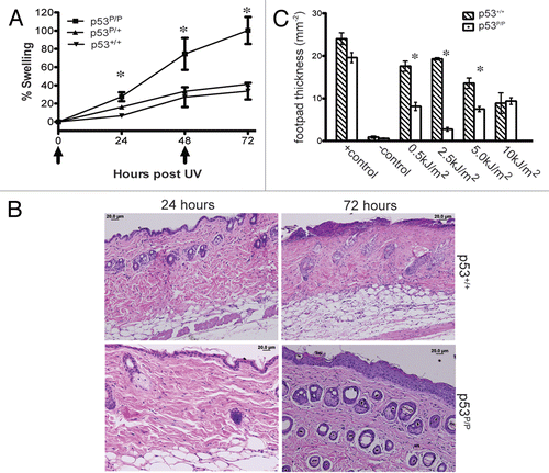
Figure 2 Induction of p53/p21 induced cellular senescence in p53R172P skin after UVB. (A) Representative skin sections from p53P/P, p53P/+ and p53+/+ mice 24 and 72 hours after 5 kJ/m2 of UVB, (top and middle panels), or 72 hours post 10 Gy of IR (bottom panels). All samples were stained with SA-β-gal followed by H&E counterstaining. Magnification, 20X. (B) Representative immunofluorescent staining of skin sections from p53P/P and p53+/+ mice; p53 (green) and p21 (red). Magnification, 40X.
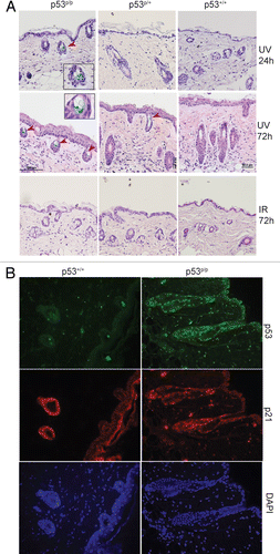
Figure 3 p53R172P MEFs are specifically hypersensitive to UVR but not IR irradiation. (A) Growth curves of p53+/+, p53P/P and p53−/− MEFs after different doses of UVB. (B) Growth curves after different doses of IR. Survival is determined by the percentage of viable cells at each treated dose over the total viable cells in untreated conditions. Representative results are presented; each experiment was repeated independently more than 5 times. Data are the mean ± SEM, *indicates p < 0.001 vs. wild-type. (C) DNA was extracted from MEFs 0, 6, 24 and 48 h post UVB exposure (200 J/m2). Photoproducts were measured by RIA using antibodies specific for CPDs or (6-4) photoproducts (6-4 PP). Representative results are presented; each experiment was repeated independently more than 3 times. (D) DNA from dorsal skin of mice 0, 6 and 24 h post UVB exposure (5 kJ/m2); photoproducts were measured as described above. N = 5 mice per time point.
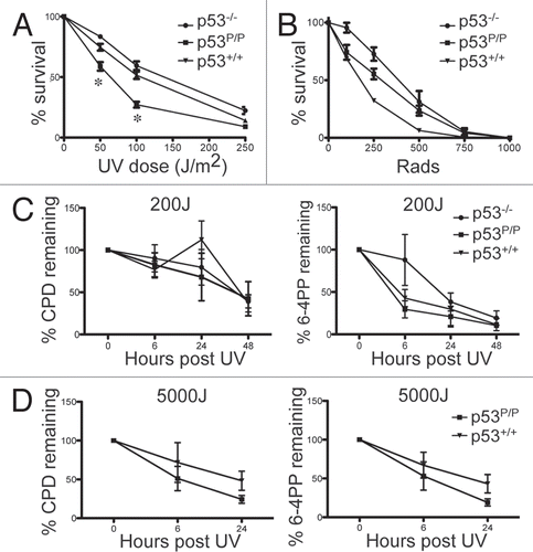
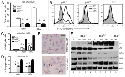
Figure 4 Wild-type MEFs undergo apoptosis while p53R172P MEFs trigger cellular senescence after UVB. (A) Cells were harvested 48 hours post UVR (100 and 250 J/m2) and apoptosis was measured using Annexin V and PI staining followed by FACS analysis. Error bars represent mean ± SEM. Differences determined using a two-tailed, unpaired student t-test. *indicates p < 0.001 vs. wild-type. (B) FACS profiles after Annexin V staining from different genotypes 48 h after UVR (100 and 250 J/m2). Representative results are presented; each experiment was repeated independently 3 times. (C) Quantification of SA-β-gal positive cells 48 h after UVB (100 and 250 J/m2). (D) Quantification of SA-β-gal 48 h after IR (6 and 10 Gy). Error bars represent mean ± SEM. *indicates p < 0.005 vs. wild-type. (e) p53P/P MEFs, untreated (upper part) and those exposed to 100 J/m2 of UVB radiation (lower part) were stained with SA-β-gal and counterstained with H&E. Magnification 40x. (F) Western blot analyses of p53, p21, PUMA, Noxa and Bcl-2 in p53+/+ and p53P/P MEFs that were exposed to 100 J/m2 of UVB and harvested 24, 48 and 72 h post irradiation, using untreated p53−/− MeFs as a control, β-actin is shown as a loading control.