
Abstract
Pura is a nucleic acid-binding protein with DNA-unwinding activity, which has recently been shown to have a role in the cellular response to DNA damage. We have investigated the function of Pura in Ultraviolet-C (UVC) radiation-induced DNA damage and nucleotide excision repair (NER). Mouse embryo fibroblasts from PURA-/- knockout mice, which lack Pura, showed enhanced sensitivity to UVC irradiation as assessed by assays for cell viability and clonogenicity compared to Pura positive control cultures. In reporter plasmid reactivation assays to measure the removal of DNA adducts induced in vitro by UVC, the Pura-negative cells were less efficient in DNA damage repair. Pura-negative cells were also more sensitive to UVC-induced DNA damage measured by Comet assay and showed a decreased ability to remove UVC-induced cyclobutane pyrimidine dimers. In wild-type mouse fibroblasts, expression of Pura is induced following S-phase checkpoint activation by UVC in a similar manner to the NER factor TFIIH. Moreover, co-immunoprecipitation experiments showed that Pura physically associates with TFIIH. Thus, Pura has a role in NER and the repair of UVC-induced DNA damage.
Acknowledgements
We thank past and present members of the Center for Neurovirology for their insightful discussion and sharing of ideas and reagents especially Dr. Nune Darbinian. We also wish to thank C. Schriver for editorial assistance. This work was supported by grants awarded by the NIH to M.K.W., E.J. and K.K.
Figures and Tables
Figure 1 Cell viability and clonogenicity of Purα-positive (Purα+) and Purα-negative (Purα−) cells in response to UVC treatment. (A) Purα+ and Purα− MEFs were treated with different doses of UVC and assayed for viability after 96 h by MTT assay. Values obtained for each UVC dose were normalized relative to UVC-untreated Purα+ cells. ■-Purα+; □-Purα+. (B) Purα+ and Purα− MEFs were treated with 10 J/m2 UVC and MTT assays performed at the time points indicated. Values were normalized relative to UVC-untreated cells at 24 h. ■-Purα+ untreated; ▴-Purα+ treated with 10 J/m2 UVC; □-Purα− untreated;. ▵-Purα− treated with 10 J/m2. (C) Purα+ and Purα− MEFs were transfected with non-target siRNA or siRNA specific for Purα at 24 h before UVC treatment (10 J/m2) and MTT assays were performed after 96 h. Values were normalized relative to UVC-untreated cells at 24 h. ■-Purα+ + NT siRNA-UVC; □-Purα+ + Purα si RNA-UVC; ▴-Purα+ + NT siRNA + UVC; ▵-Purα+ + Purα si RNA + UVC. (D) Purα+ and Purα− MEFs were transfected with pCMV plasmid or pCMV-Purα expression plasmid, treated with different doses of UVC and assayed for viability after 96 h by MTT assay. Values were normalized to UVC-untreated MEF Purα− (+pCMV) cells. ■-Purα+ + pCMV-Purα; □-Purα+ + pCMV. (E) Western blot for Purα for the cells used in (A and B) with α-tubulin as a loading control. (F) Western blot for Purα for the cells used in (C) with α-tubulin as a loading control. (G) Western blot for Purα for the cells used in (D) with α-tubulin as a loading control. (H) Purα+ and Purα− MEFs were treated with UVC as indicated and plated. After two weeks, colonies were counted and values normalized relative to unirradiated controls. The experiments were repeated five times. *p < 0.03.
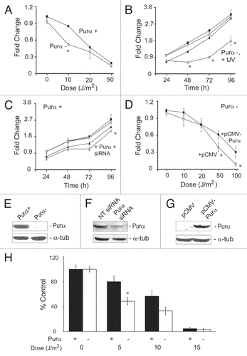
Figure 2 Reactivation of transfected UVC-irradiated reporter plasmid in Purα+ and Purα− cells. Luciferase reporter plasmid was treated in vitro with and without UVC as described in Materials and Methods and then introduced into Purα+ and Purα− cells by transfection. (A) Dose-response for restoration of luciferase activity. (B) In this experiment, expression vectors pCMV-Purα and pCMV were included in the transfections together with the irradiated plasmid. The experiments were repeated at least three times. *p < 0.03.
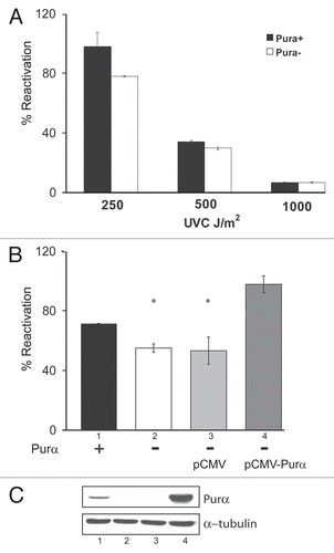
Figure 3 Assays of cyclobutane pyrimidine dimers (CPDs) in UV-irradiated Purα+ and Purα− cells. (A) Purα+ and Purα− cells were synchronized by 48 h serum deprivation and treated with 10 J/m2 of UVC. After 24 h or at time zero, cells were fixed with 70% ethanol and labeled for CPDs with anti-thymine antibody and FITC-conjugated secondary antibody. After adding propidium iodide, G1/G0 cell populations were analyzed by flow cytometry for CPDs. Histograms show fractions of CPD-positive cells in green in the analyzed samples. (B) Purα+ and Purα− MEFs were synchronized in G1/G0, treated with UVC, DNA isolated and analyzed by slot immunoblotting using monoclonal antibody against CPD as described in Materials and Methods. The densities of the bands were quantified by densitometry and the graphs were plotted after normalization to the amount of loaded DNA. The experiments were repeated at least three times. *p < 0.05.
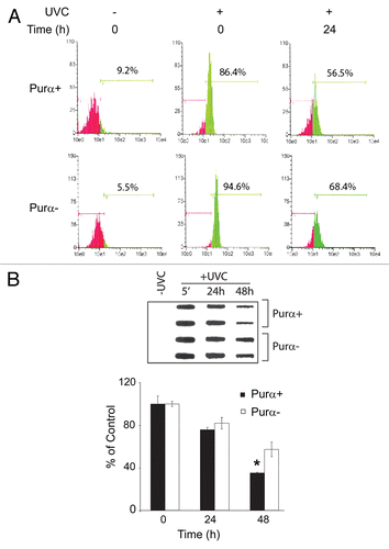
Figure 4 DNA fragmentation measured by the Comet assay in UV-irradiated Purα+ and Purα− cells. (A) Purα+ and Purα− cells were synchronized by 48 h of serum deprivation, treated with 50 J/m2 of UVC irradiation, subject to in situ electrophoresis and the tail of fragmented DNA measured (Olive Tail Moment) as described in Materials and Methods (Comet assay). The OTM was scored randomly from 100 propidium iodide labeled cells, for each condition. The plot shows a percentage of the Comet positive nuclei (OTM>13), apoptotic and necrotic cells were not scored. (B) Representative photomicrograph of cells with low and high OTM. The experiments were repeated at least three times.
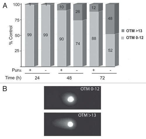
Figure 5 Effect of UVC on DNA damage signaling pathways in Purα+ and Purα− cells. Purα+ and Purα− cells were synchronized by double thymidine block (G1/S block, 2 mM thymidine) and treated with 50 J/m2 of UVC. After 6 and 24 h, whole cell protein extracts were analyzed by western blot.
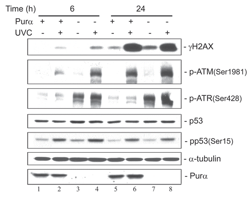
Figure 6 Effect of UVC irradiation on synchronized wild-type Balb/c mouse fibroblasts. Wild-type Balb/c mouse fibroblasts were synchronized by 72 hours of serum starvation and then treated with and without UVC (20 J/m2) at time zero. (A) Cells were harvested and subject to western blot. (B) The western blots in (A) were quantitated by densitometry. (C) Cells from the same samples were harvested and analyzed by FACS for the cell cycle.
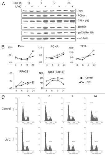
Figure 7 Co-immunoprecipitation of GFP-Purα with TFIIH. Purα− MEFs were transfected with expression plasmid for GFP or GFP-Purα and whole cell extracts prepared. Immuno-precipitation was performed with antibody for TFIIH/p89 (lanes 3 and 4) or nonimmune rabbit serum (NRS-lane 5) and the immune complex analyzed by western blot using antibody to GFP. Lanes 1 and 2 shows 1/10 of the immunoprecipitation input from the GFP- and GFP-Purα−transfected whole cell extracts respectively.
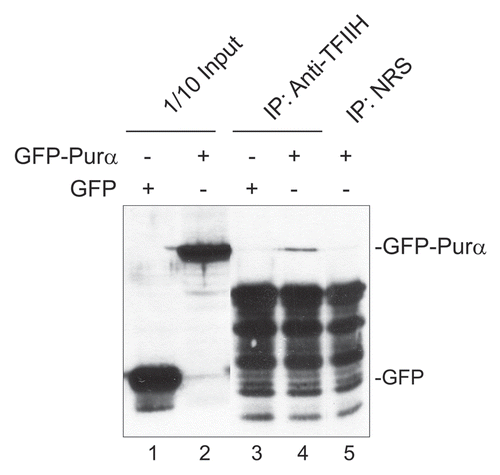
Table 1 Cell cycle profiles of Purα+ and Purα− cells with or without UVC irradiation