
Abstract
Hereditary leiomyomatosis renal cell carcinoma (HLRCC)-associated renal tumors are aggressive and tend to metastasize early. There are currently no effective forms of therapy for patients with advanced HLRCC-associated kidney cancer. We have previously shown that HLRCC cells express a high level of reactive oxygen species (ROS). In the present study we investigated the cytotoxiceffects of increasing ROS level using bortezomib in combination with cisplatin on HLRCC cells in vitro and in an in vivo xenograft model. The cytotoxic effect of several ROS inducers on FH-deficient cells was assessed by synthetic lethality. ROS inducers had a pronounced impact on the viability of FH-deficient cells. Because of its high potency, the proteasome inhibitor bortezomib was further investigated. Bortezomib induced apoptosis in vitro in HLRCC cells and inhibited HLRCC tumour growth in vivo. Bortezomib-associated cytotoxicity was highly correlated with cellular ROS level: combining bortezomib with other ROS inducers enhanced cytotoxicity, while combining bortezomib with a ROS scavenger inhibited its cytotoxic effect. Finally, HLRCC murine xenografts were treated with bortezomib and cisplatin, another ROS inducer. This regimen induced HLRCC tumour regression in vivo. These findings suggest that increasing ROS level in HLRCC above a certain threshold can induce HLRCC-tumor cell death. Increasing tumor ROS with bortezomib in combination with cisplatin represents a novel targeted therapeutic approach to treat advanced HLRCC-associated renal tumors.
Acknowledgements
We wish to thank Dr. B.T. Scroggins for helpful discussions, Ms. Catherine Wells for technical assistance and Ms. G. Shaw for editorial assistance. This research was supported by the Intramural Research Program of the NIH, National Cancer Institute, Center for Cancer Research.
Figures and Tables
Figure 1 The proteasome inhibitor bortezomib induces apoptosis of HLRCC cells in vitro and in vivo. (A) The apoptosis marker cleaved caspase 3 was visualized by western blot. (B) Cell viability of UOK262 treated with the pan-caspase inhibitor Z-VAD (10 uM) 1 h prior to bortezomib (5 nM, 16 h). (C) Scid/beige mice were treated with bortezomib (1 mg/kg, twice weekly) for 3 weeks. Tumor volumes were measured with a caliper prior to each treatment. Arrows show when the mice were treated. (D) TdT staining was performed on five-micron slides from formalin-fixed, paraffin embedded HLRCC xenograft samples. Z-VAD: Z-VAD-FMK; * and #p < 0.05.
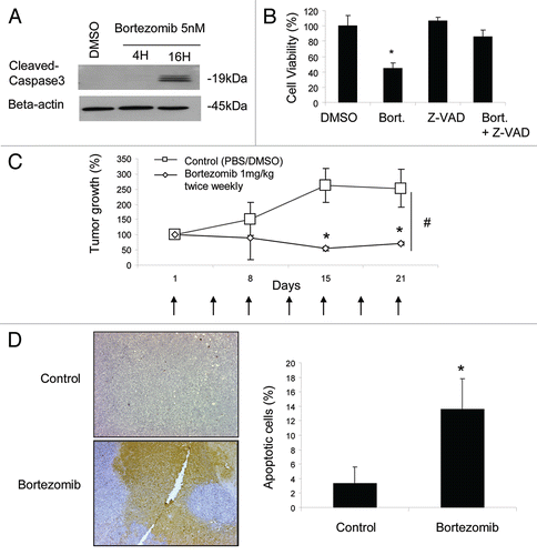
Figure 2 Bortezomib-induced toxicity correlates with ROS level in FH-deficient cells. (A) ROS level was measured with H2DCFDA after treatment for 4 h with bortezomib (bort., 5 nM), menadione (Mena., 6 uM), and/or n-acetyl-cysteine (NAC, 5 mM). (B) Viability of UOK262 cells after bortezomib (bort., 5 nM) treatment alone or in combination with ROS modulators (Mena: menadione, 6 uM; NAC, 5 mM); * and #p < 0.05.
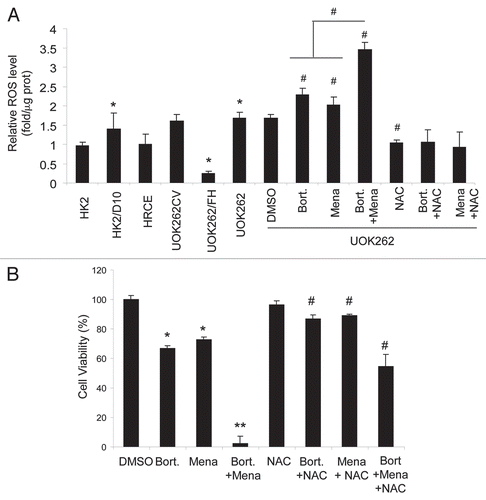
Figure 3 Bortezomib's effect is enhanced in vitro by the ROS inducer cisplatin. (A) ROS level was measured with H2DCFDA after treatment for 4 h with bortezomib (bort., 5 nM), cisplatin (Cispl., 6uM), and n-acetyl-cysteine (NAC, 5 mM) in UOK262 cells. (B) Cell viability in UOK262 and HRCE cells 24 h after treatment with bortezomib (bort., 5 nM), cisplatin (Cispl., 6 uM), and n-acetyl-cysteine (NAC, 5 mM), alone or in combination. * and #p < 0.05.
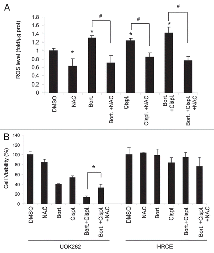
Figure 4 Combining cisplatin and bortezomib induces HLRCC tumour regression in vivo. (A) Scid/beige mice were treated i.p. once weekly with bortezomib (0.1 mg/kg) and cisplatin (2.5 mg/kg), alone or in combination. Arrows show when the mice where treated. (B) Five-micron slides from formalin-fixed, paraffin embedded samples were utilized for TdT staining. # and *p < 0.05.
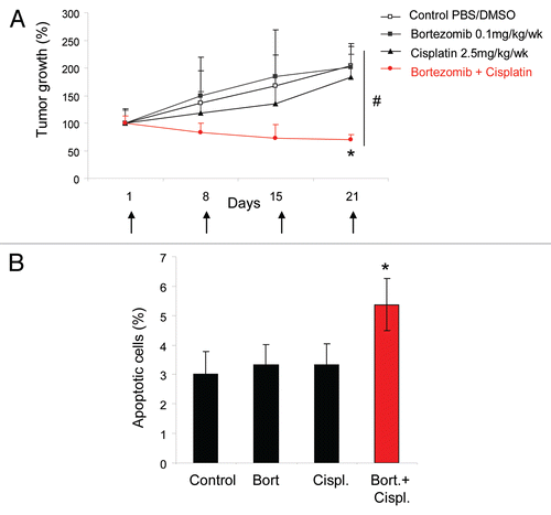
Table 1 Cytotoxicity of ROS-inducing agents correlates inversely with fumarate hydratase activity status