Abstract
Activation of the DNA damage response (DDR) is critical for genomic integrity and tumor suppression. The occurrence of DNA damage quickly evokes the DDR through ATM/ATR-dependent signal transduction, which promotes DNA repair and activates the checkpoint to halt cell cycle progression (Halazonetis et al., 2008; Motoyama and Naka, 2004; Zhou and Elledge, 2000). The "turn off" process of the DDR upon satisfaction of DNA repair, also known as "checkpoint recovery", involves deactivation of DDR elements, but the mechanism is poorly understood. Greatwall kinase (Gwl) has been identified as a key element in the G2/M transition (Archambault et al., 2007; Jackson, 2006; Zhao et al., 2008; Yu et al., 2004; Yu et al., 2006; Zhao et al., 2006) and helps maintain M phase through inhibition of PP2A/B55δ (Burgess et al., 2010; Castilho et al., 2009; Goldberg, 2010; Lorca et al., 2010; Vigneron et al., 2009), the principal phosphatase for Cdk-phosphorylated substrates. Here we show that Gwl also promotes recovery from DNA damage and is itself directly inhibited by the DNA damage response (DDR). In Xenopus egg extracts, immunodepletion of Gwl increased the DDR to damaged DNA, whereas addition of wild type, but not kinase dead Gwl, inhibited the DDR. The removal of damaged DNA from egg extracts leads to recovery from checkpoint arrest and entry into mitosis, a process impaired by Gwl depletion and enhanced by Gwl over-expression. Moreover, activation of Cdk1 after the removal of damaged DNA is regulated by Gwl. Collectively, these results defines Gwl as a new regulator of the DDR, which plays an important role in recovery from DNA
Acknowledgements
This work was supported by the Howard Hughes Medical Institute and by a grant from the NIH to M.L.G. (GM48430). A.P. and T.M.M. are Research Associates and J.L.M. an Investigator of the Howard Hughes Medical Institute.
Figures and Tables
Figure 1 Gwl negatively regulates the DNA damage response. Interphase Xenopus egg extracts were mock-treated (beads alone), immunodepleted for Gwl or Gwl-depleted and then reconstituted with purified wild-type (WT) or kinase-dead (KD) Gwl, prepared as described previously.Citation15 Extracts were then supplemented with double-stranded oligonucleotides (dA-dT) at 20 ug/ml, incubated for 30 min at room temperature and analyzed for the DDR by western blotting using the indicated antibodies.
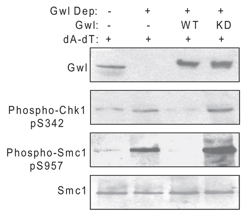
Figure 2 DNA damage inhibits Gwl activity. (A) Interphase Xenopus egg extracts with or without dA-dT (20 ug/ml) were incubated at room temperature for 15 (−), 30 or 90 min, as indicated. Fifteen min before harvest, the extract was transferred to a tube containing a FLAG-Gwl bead pellet corresponding to the amount of Gwl expressed in three oocytes. At the end of the incubation, Gwl beads were spun down and used for kinase assay as described in Materials and Methods. The supernatant was supplemented with SDS-PAGE sample buffer and then analyzed by western blotting for phospho-Smc1, Smc1 and phospho-Cdc2. An autoradiograph of phosphorylated MBP is shown. (B) Interphase egg extracts were supplemented with or without dA-dT and caffeine (10 mM) as indicated for 10 min. Then extract was added to a tube containing a FLAG-Gwl bead pellet corresponding to the amount of Gwl expressed in three oocytes. After a further 20 min incubation, the beads were re-isolated by centrifugation and used for determination of Gwl kinase activity as described in Materials and Methods. The supernatant was analyzed by western blotting for phospho-Smc1 and Smc1. Equal loading in the immune-complex kinase assay was verified by western blotting of the assay for FLAG-Gwl and by Coomassie staining of the assay substrate (MBP). Liquid scintillation counting of the excised MBP bands indicates approximately 80% inhibition of Gwl activity after 30 min of DNA damage (data not shown).
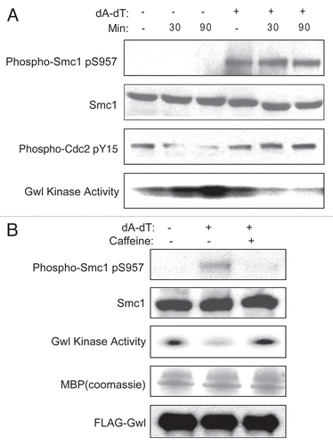
Figure 3 Recovery from the DNA damage response in extracts. (A) The cell-free checkpoint recovery system. Metaphase II-arrested CSF extracts were released into interphase for 30 min by addition of Ca++. Magnetic beads conjugated with dA-dT were added to 20 µg/ml and incubated in the extract for 30 min then removed with a magnet as described in Materials and Methods to mimic the completion of DNA repair. (B) Analysis of dA-dT beads removed from extracts. As in (A), extracts before and after dA-dT removal, as well as the removed beads were analyzed by western blotting using the indicated antibodies. Lane 1, extract before dA-dT removal; lane 2, extract after dA-dT removal; lane 3, dA-dT beads removed from lane 1. (C) The removal of dA-dT from extracts depicted in (A) enables deactivation of DNA damage signaling and re-entry into mitosis, as judged by western blotting of Smc1, Chk1, Cdc2 and Cdc27. Control extracts without removal of dA-dT (left) sustained the checkpoint arrest.
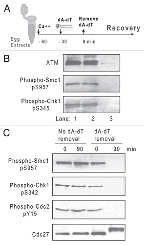
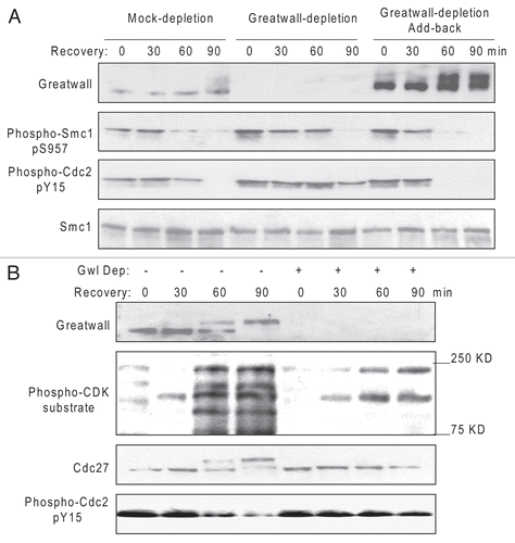
Figure 4 Gwl promotes checkpoint recovery. (A) As in , biotinylated dA-dT oligos bound to M-280 streptavidin beads were added to extracts for 30 min to activate the DNA damage checkpoint and then removed with a magnet to allow recovery. As indicated, these extracts had also been either mock-treated with Protein G Dynabeads or depleted of Gwl with anti-Gwl antibody bound to Protein G Dynabeads as described in Materials and Methods, or Gwl-depleted and then reconstituted with purified Gwl. Recovery in the extracts was then analyzed at 90 min by western blotting with the indicated antibodies. (B) As in (A) extracts with or without depletion of Gwl were monitored for Cdk activation by western blotting with the indicated antibodies at various time points during recovery.