Abstract
Understanding the regulatory mechanisms controlling the fate decisions of neural stem cells (NSCs) is a crucial issue to shed new light on mammalian central nervous system (CNS) development in health and disease. We have investigated a possible role for the previously uncharacterized BTB/POZ-domain containing zinc finger factor Zbtb45 in the differentiation of NSCs and postnatal oligodendrocyte precursors. In situ hybridization histochemistry and RT-qPCR analysis revealed that Zbtb45 mRNA was ubiquitously expressed in the developing CNS in mouse embryos at embryonic day (E) 12.5 and 14.5. Zbtb45 mRNA knockdown in embryonic forebrain NSCs by siRNA resulted in a rapid decrease in the expression of oligodendrocyte-characteristic genes after mitogen (FGF2) withdrawal, whereas the expression of astrocyte-associated genes such as CD44 and GFAP increased compared to control. Accordingly, the number of astrocytes was significantly increased seven days after Zbtb45 siRNA delivery to NSCs, in contrast to the numbers of neuronal and oligodendrocyte-like cells. Surprisingly, mRNA knockdown of the Zbtb45-associated factor Med31, a subunit of the Mediator complex, did not result in any detectable effect on NSC differentiation. Similar to NSCs, Zbtb45 mRNA knockdown in oligodendrocyte precursors (CG-4) reduced oligodendrocyte maturation upon mitogen withdrawal associated with down-regulation of the mRNA expression and protein levels of markers for oligodendrocytic differentiation. Zbtb45 mRNA knockdown did not significantly affect proliferation or cell death in any of the cell types. Based on these observations, we propose that Zbtb45 is a novel regulator of glial differentiation.
Acknowledgements
We would like to thank Dr. J.C. Louis (Amgen) for the CG4 cell line, Stephanie Robertson, Gon#x000E7;alo Castelo-Branco, Anna Cascante, and other members of the Hermanson lab for input, and Christian Broberger and Kylie Foo for valuable technical advice. This study was supported by grants from the Swedish Research Council (VR), the Swedish Cancer Society (CF), the Jeansson Foundation, the Åke Wiberg Foundation, the Åhlén Foundation, the Swedish Medical Society, Karolinska Institutet, the Swedish Foundation for Strategic Research (SSF), K&A Wallenberg Foundation (CLICK), and the Swedish Childhood Cancer Foundation (BCF) to O.H.
Figures and Tables
Figure 1 The predicted Zbtb45 amino acid sequence displayed typical features of a Zbtb protein. (A) Alignment of the predicted amino acid sequence of Zbtb45 to Zbtb20, Zbtb7, PLZF and Bcl-6 confirmed that the N-terminal part of the putative protein contained highly conserved residues that contribute to protein stability (black triangles) as well as highly conserved components of a charged pocket that is required for the function of Zbtb proteins (framed). (B) Phylogenetic tree showing that the predicted Zbtb45 amino acid sequence share closest homology to Zbtb20. (C) The Zbtb45 gene was predicted to be encoded into a 520-residue transcript with a BTB/POZ domain proximal to the N terminus and four predicted zinc fingers of C2H2-type in the C terminus.
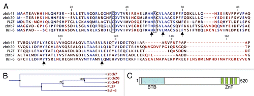
Figure 2 Ubiquitous expression of Zbtb45 mRNA in the developing mouse forebrain. (A and C) In situ hybridization histochemistry of coronal brain sections from embryonic mice revealed that Zbtb45 mRNA was present in the developing forebrain at (A) E12.5 and (C) E14.5. (B and D) DAPI staining on adjacent and same sections. (E) The ubiquitous expression pattern was further confirmed by qPCR analysis of Zbtb45 mRNA levels relative to TATA-binding protein in rat E15 cortex (neocortex; NCx), ganglionic eminences (GE; lateral GE—LGE, medial GE—MGE), thalamus (Th), olfactory bulb (OB), heart and forelimb (Limb), showing only minor regional variations. (F) RT-qPCR analysis of neural stem cells (NSCs) revealed that FGF2 withdrawal had little effect on the expression of Zbtb45 at 1 and 2 DIV. Bars represent the levels of Zbtb45 in FGF2-conditions relative to the levels in FGF2+ conditions. Results from representative experiments are shown (A–F).
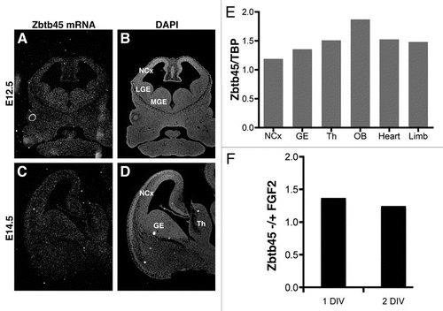
Figure 3 Zbtb45 siRNA delivery to NSCs yielded increased astrocyte- and decreased oligodendrocyte gene expression. (A) siZbtb45 (right part) lead to an increased number of astrocytes after 7 DIV compared to control (left part). Immunocytochemistry with GFAP antibody (green) and MBP (red) with DAPI (blue) counterstain. 20x magnification. (B) NSC cultures lacking Zbtb45 contained significantly increased numbers of GFAP+ cells, whereas numbers of MBP+ and TuJ1 cells were unaffected compared to control. (C–E) RT-qPCR analysis of mRNA levels in NSCs after 1 (gray bars) and 2 DIV (black bars) revealed that siRNA against Zbtb45 led to significant changes in expression levels of markers for astrocytes and oligodendrocytes but only moderate changes in gene expression of various transcription and signaling factors. Bars indicate mRNA levels of siZbtb45 cells relative to control. (F) RT-qPCR analysis of mRNA levels in NSCs after 1 (gray bar) and 2 DIV (black bar) suggested that siRNA mediated knockdown of the Zbtb45-associated factor Med31 had no effect on differentiation. n = 3 (B) or 5 (C–F) independent experiments (*p < 0.05, **p < 0.01), error bars = SEM.
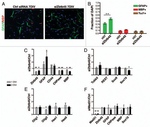
Figure 4 Delivery of Zbtb45 siRNA to CG-4 oligodendrocyte precursors inhibited oligodendrocyte maturation. (A) RT-qPCR analysis of mRNA levels in NSCs after 1 (gray bars) and 3 DIV (black bars) revealed that siRNA against Zbtb45 led to significant changes in expression levels of markers oligodendrocytes along with an upregulation of the astrocyte related gene CD44. Bars indicate mRNA levels of siZbtb45 cells relative to control. (B) CG-4 cultures lacking Zbtb45 contained significantly less MBP+ cells. (C and D) Immunocytochemistry with MBP (red) with DAPI (blue) counterstain. 20x magnification. siZbtb45 lead to a marked decrease in mature oligodendrocytes after 7 DIV compared to control. n = 3 independent experiments (**p < 0.01), error bars = SEM.
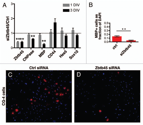
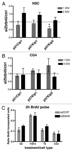
Figure 5 Zbtb45 siRNA did not significantly affect proliferation or cell cycle inhibitor expression. (A) RT-qPCR analysis of mRNA levels of cyclin-dependent kinase inhibitors in NSCs 1 and 2 DIV after Zbtb45 knockdown. A temporal, significant downregulation of p57kip2 mRNA levels was observed at 1 DIV (B) RT-qPCR analysis of mRNA levels of cyclin-dependent kinase inhibitors in CG-4 cells 1 and 3 DIV after Zbtb45 knockdown. (C) Bars indicate numbers of cells incorporating BrdU after a 2 h pulse relative to total number of nuclei. No significant difference in BrdU incorporation was detected in NSCs lacking Zbtb45 relative to control in N2 media, presence of FGF2 or thyroid hormone (T3) or CG-4 cells in proliferating conditions. n = 5 (A) or 3 (B and C) independent experiments (*p < 0.05), error bars = SEM.