Abstract
Smad3, a component of the TGFβ signaling pathway, contributes to G1 arrest in breast cancer cells. Overexpression of the cell cycle mitogen, cyclin E, is associated with poor prognosis in breast cancer, and cyclin E/CDK2 mediated phosphorylation of Smad3 has been linked with inhibition of Smad3 activity. We hypothesized that the biological aggressiveness of cyclin E overexpressing breast cancer cells would be associated with CDK2 phosphorylation and inhibition of the tumor suppressant action of Smad3. Expression constructs containing empty vector, wild type (WT) Smad3, or Smad3 with CDK phosphorylation site mutations were co-transfected with a Smad3-responsive reporter construct into parental, vector control (A1), or cyclin E overexpressing (EL1) MCF7 cells. Smad3 function was evaluated by luciferase reporter assay and mRNA analysis. The impact of a Cdk2 inhibitor and cdk2 siRNA on Smad3 activity was also assessed. Cells expressing Smad3 containing mutations of the CDK phosphorylation sites had higher p15 and p21 and lower c-myc mRNA levels, as well as higher Smad3-responsive reporter activity, compared with controls or cells expressing WT Smad3. Transfection of cdk2 siRNA resulted in a significant increase in Smad3-responsive reporter activity compared with control siRNA; reporter activity was also increased after the treatment with a Cdk2 inhibitor. Thus, cyclin E-mediated inhibition of Smad3 is regulated by CDK2 phosphorylation of the Smad3 protein in MCF7 cells. Inhibition of CDK2 may lead to restoration of Smad3 tumor suppressor activity in breast cancer cells, and may represent a potential treatment approach for cyclin E overexpressing breast cancers.
Acknowledgements
We thank Dr. Fang Liu (Rutgers, State University of New Jersey, Piscataway, NJ) for providing the mutant Smad3 constructs, Dr. Khandan Keyomarsi (MD Anderson Cancer Center, Houston, TX) for providing the A1 control and EL1 cyclin E overexpressing MCF7 cells, and Elizabeth Tarasewicz, A. June Gordon, and Dr. Stacey C. Tobin for editorial assistance.
Financial Support
J.S.J. is a Lynn Sage Scholar supported by the American Cancer Society-Illinois Division, the Dixon Foundation, the Association of Women Surgeons and an NIH K22 CA138776 research grant.
Figures and Tables
Figure 1 Characterization of MCF7 stably transfected cells. (A) Protein expression levels were measured in extracts from parental cells (MCF7) or MCF7 clones stably transfected with empty vector (A1) or cyclin E (EL1). Total protein (30 µg/lane) was separated on an SDS/PAGE gel and subjected to immunoblot analysis with the indicated antibodies. (B) CDK2 activity in cyclin E overexpressing MCF7 cells. CDK2 kinase assay radiographs and densitometric quantification in MCF7 study cell lines (parental, MCF7; vector control, A1; cyclin E overexpressing, EL1). CDK2 kinase activity was quantified relative to the amount of activity in the parental line.
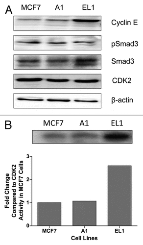
Figure 2 Localization of Smad3 in the MCF7 study cell lines. Study cell lines underwent immunofluorescent staining to detect Smad3. DAPI indicates nuclei, FITC indicates Smad3 protein. The overlay demonstrates that Smad3 is localized to both the nuclear and cytoplasmic compartments (arrowheads) in the parental, vector control (A1) and cyclin E overexpressing (EL1) cell lines.
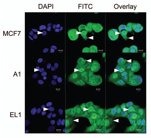
Figure 3 Expression of Smad3-regulated genes in study cell lines. MCF7 study cells were transfected with wild-type Smad3 (WT) or the 5M Smad3 CDK phosphorylation site mutant and transcript levels of c-myc, p15 and p21 were measured by real-time quantitative RT-PCR. * denotes significant difference from cells transfected with CS2 control for each cell line.
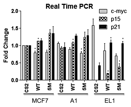
Figure 4 Relative transcriptional activity of various Smad3 constructs in cyclin E overexpressing MCF7 cells. Cells were co-transfected with the Smad3-responsive CAGA-luc reporter construct and Renilla luciferase reporter, in addition to the indicated Smad3 expression constructs. Data are shown as fold increase in normalized luciferase activity (firefly/Renilla) compared with empty vector-transfected MCF7 cells (CS2). Error bars indicate standard deviation from the mean of normalized luciferase activity for each study condition. * denotes significant difference from cells transfected with the WT Smad3 for each cell line.
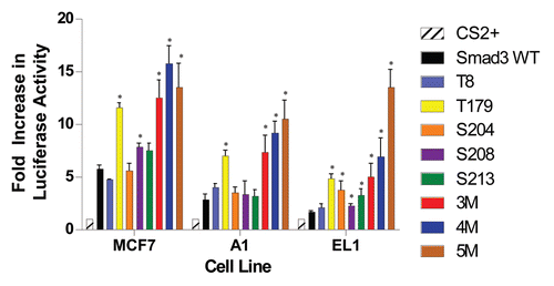
Figure 5 Effect of CDK2 inhibition on Smad3 transcriptional activity in MCF7 cells. (A) Dose-dependent increase in Smad3 transcriptional activity in MCF7 cells transfected with the Smad-responsive CAGA-luc reporter construct, Renilla luciferase reporter and wild-type (WT) Smad3 expression vector, then treated with the indicated concentrations of CDK2 inhibitor (CDK2i). (B) Restoration of Smad3 transcriptional activity with inhibition of CDK4 activity. MCF7 study cell lines were transfected with the Smad3-responsive CAGA-luc reporter construct, Renilla luciferase reporter, and either wild-type Smad3 (WT), T179 Smad3 (T179) or 5M Smad3 (5M) expression vectors. The cells were treated with vehicle or 240 nM CDK2 inhibitor (CDK2i). Data are shown as fold increase in normalized luciferase activity (firefly/Renilla) compared with empty vector-transfected MCF7 cells (CS2). Error bars indicate standard deviation from the mean of normalized luciferase activity for each study condition. * denotes significant difference from cells transfected with WT Smad3 for each cell line.
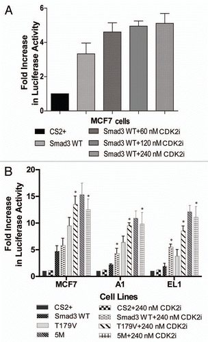
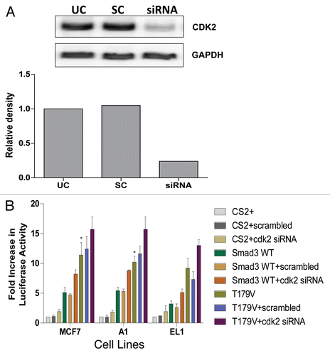
Figure 6 Restoration of Smad3 transcriptional activity with siRNA knockdown of CDK2. (A) MCF7 cells were transfected with scrambled (SC) or cdk2 siRNA (siRNA) for 48 h, then the cells were lysed and the level of CDK2 protein was determined in untransfected cells (UC) and transfected cells by immunoblot analysis using anti-CDK2 antibody. GAPDH was used as a loading control. (B) MCF7 study cell lines were co-transfected with the Smad3-responsive CAGA-luc reporter construct and Renilla luciferase reporter; siRNA or cdk2 siRNA; and either empty vector (CS2), wild-type Smad3 (WT) or T179 Smad3 mutant (T179) expression vectors. Data are shown as fold increase in normalized luciferase activity (firefly/Renilla) compared with empty vector-transfected MCF7 cells. Error bars indicate standard deviation from the mean of normalized luciferase activity for each study condition. * denotes significant difference from cells transfected with WT Smad3 and scrambled siRNA for each cell line.