
Abstract
Accurate chromosome segregation during mitosis is achieved by the kinetochore fibres (K-fibres) of the spindle apparatus. These fibers are bundles of microtubules (MTs) connected by non-motor bridges. We recently identified a TACC3/ch-TOG/clathrin complex that constitutes the shortest class of inter-MT bridge in K-fibers. TACC3 anchors the complex to MTs and this is dependent on phosphorylation by Aurora A kinase. Here we show that inhibition of Aurora A kinase using MLN8237 results in 1) loss of clathrin and TACC3 from spindles, 2) destabilization of K-fibers and 3) loss of inter-MT bridges. These results are similar to those in cells depleted of clathrin or TACC3; suggesting that TACC3/ch-TOG/clathrin bridges are the major class of bridge that is regulated by this kinase.
Acknowledgements
We thank Bill Earnshaw for the gift of anti-CENP-B. This work was supported by a Career Establishment Award from Cancer Research UK (C25425/A8722). L.P.C. and D.G.B. are recipients of Wellcome Trust Prize Studentships and I.A.P. is a Royal Society University Research Fellow.
Figures and Tables
Figure 1 Effect of inhibition of Aurora A kinase on TACC3/clathrin localization and K-fiber stability. (A) Inhibition of Aurora A kinase resulted in loss of clathrin and TACC3 from mitotic spindles. HEK293 cells near metaphase were treated with MLN8237 (0.5 µM, 40 min) prior to fixation and staining with the indicated antibodies. All experimental details are as described previously in reference Citation7, anti-NuMA (#3888, Cell Signaling). Bar, 10 µm. (B) Inhibition of Aurora A kinase resulted in destabilization of kinetochore fibers. HeLa cells near metaphase were treated with no drug (Control) or MLN8237 (0.3 µM, 40 min) and then incubated for 6 min in warm (control) or cold (cold treated) media to depolymerize any non-stable MTs. Cells were fixed and stained for βtubulin (green), CENP-B (red) and DNA (DAPI, blue). Bar, 10 µm. Note the misaligned chromosomes (arrows) and the “orphan” centromeres in the inset. Similar observations were reported in clathrin-depleted cells.Citation9,Citation10
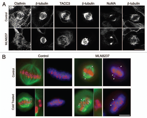
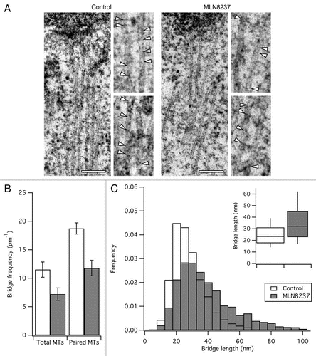
Figure 2 Effect of inhibition of Aurora A kinase on inter-MT bridges. Inhibition of Aurora A kinase resulted in loss of inter-MT bridges. MLN8237-treated HeLa cells near metaphase were processed for EM. (A) Representative sections to show the morphology of K-fibers. Zoomed regions show bridges indicated by arrowheads. Scale bar, 200 nm. (B and C) Quantification of inter-MT bridges in K-fibers from control or MLN-treated cells. (B) Bar chart to show the average frequency of bridges per unit length of total or paired MTs. Student's t-test, p = 0.066 and p = 0.014; Ncell = 3; Bars, mean ± SEM. (C) Frequency histogram to show the length distribution of inter-MT bridges. Inset: Tukey plots of the length distribution data.
Addendum to: