
Abstract
The corneal epithelium generates a significant trans-epithelial potential (TEP) which aids in maintaining cornea water balance and transparency. Injury to the cornea causes a short circuit of the TEP at the wound. The TEP in the intact epithelium around the wound acts like a battery, powering significant ion flux and electric current at the wound. These circulating endogenous currents generate an electric field orientated towards the wound, with the wound the cathode. Many cell types, including human corneal epithelial cells and keratinocytes, migrate to the cathode at physiological electric field strengths. Indeed, the electric signal is a powerful stimulator of cell migration which appears to over-ride other cues such as chemotaxis and wound void. These wound fields also have a dynamic timecourse of change after wounding. It has been assumed that wound electric fields are produced by passive leakage of ions from damaged cells and tissue. Could these fields be actively maintained and regulated as an active wound response? What are the molecular, ionic and cellular mechanisms underlying the wound electric currents?
Acknowledgments
This work was supported by the National Institutes of Health National Eye Institute grant 1R01EY019101 (to M.Z. and B.R.). The authors thank the Wellcome Trust for continuous support (068012). This work was also supported in part by Research to Prevent Blindness, Inc., an NSFC grant (30628026), and U.C. Davis Dermatology Department developmental fund. M.Z. is also supported by grants from the California Institute of Regenerative Medicine RB1-01417, NSF MCB-0951199.
Figures and Tables
Figure 1 Which ions contribute to the wound electric current? The corneal epithelium transports Na+ and Cl− to generate and maintain a transepithelial potential difference (TEP). Injury breaks the epithelial barrier and collapses the potential at the wound (left). The positive potential in the surrounding intact epithlium drives ion current flow out of the wound (blue arrows) and forms laterally-orientated wound electric fields (red arrow) with the wound the cathode. These fields initiate and promote healing by stimulating cell migration into the wound.
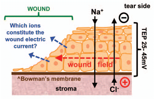
Figure 2 Relative contributions of ions to wound current. (A) Chloride channel blocker DIDS significantly reduces cornea wound electric current (*p < 0.03). (B) Overlaying wound electric current data (red) with compiled ion flux data (flux of all ions combined; blue) shows that electric current increase after wounding is due to an increasing ion flux (mainly chloride). Wound electric current data from Reid et al.Citation3 (C) Chloride is the major ion contributing to the normal (“Control”; blue) wound current. Enhancement of wound current by aminophylline (“Amin.”; red) is mostly due to stimulation of chloride flux, but also partly by reversal of sodium flux (inward to outward).
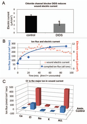
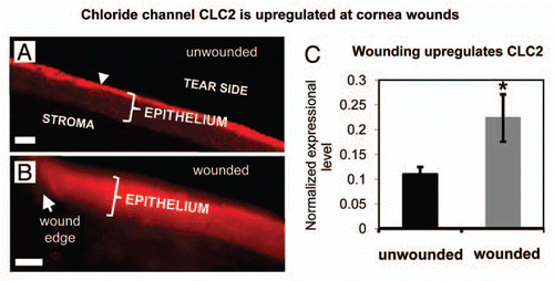
Figure 3 Distribution and expression of calcium-activated chloride channel-2 (CLC2). (A) In unwounded cornea, CLC2 channels were concentrated in the superficial epithelial cells (arrowhead). (B) One h after wounding, fluorescence was present throughout the entire thickness of the epithelium, showing re-distribution and increased concentration of CLC2 channels. Scale bars 50 µm. (C) In human corneal epithelial cell monolayer, scratch wounding induced increased expression of CLC2 channel mRN A (*p < 0.05).
Addendum to: