
Abstract
Mammalian spermatozoa become competent for fusion with oocytes while traveling through the female reproductive tract and the oocyte’s extracellular investments. Recent studies highlighted the molecular mechanism of the sperm’s interactions with the zona pellucida (ZP), the extracellular coat surrounding the oocyte. Fertilizing spermatozoa initiate the sperm acrosome reaction (AR), essential for zona penetration and fusion with the oocyte plasma membrane, before they reach the ZP. However, the exact condition of spermatozoa that leads to successful penetration of the ZP remains unknown. We performed microscopic observations of in vitro fertilization with genetically (EGFP) and chemically (antibody and lectin) labeled spermatozoa to monitor the progression of the AR. Spermatozoa exhibiting EGFP-/PNA+ prior to binding to the ZP initiated zona penetration. This result suggests that spermatozoa that have undergone the AR are still capable of binding and penetrating the ZP.
Acknowledgments
We thank Dr. Vic Vacquier for his critical reading of the manuscript. This study was supported by the Japan Society for the Promotion of Science (N.H.).
Figures and Tables
Figure 1 Histochemical dissection of the acrosomal exocytosis. (A) The diagram shows the temporal correlation between progression of the acrosomal exocytosis and the surface exposure of intra-acrosomal markers suggested by this study. An intra-acrosomal protein, sp56/ZP3R is exposed on the sperm surface before initiation of the acrosomal exocytosis as judged by the presence of soluble acrosomal EGFP fluorescence. Whereas, peanut agglutinin lectin (PNA)-reactive carbohydrate residues, that locate inside the acrosome, is exposed to the apical ridge subdomain (white arrowhead) after disappearance of EGFP fluorescence, followed by extended staining to the equatorial segment (yellow arrowhead) of sperm head, which is regarded as completion of the acrosomal exocytosis.Citation13 Although such equatorial staining was clearly seen under a fluorescence microscope, neither flow cytometry analysis nor PNA-MIVF assay could distinguish between early and late stages of PNA distributions (see below). Mitochondria were labeled with Ds-Red2. (B) Staining of transgenic sperm with Qdot® 800-labeled PNA revealed that PNA+ and EGFP+ sperm were mutually reciprocal. B/G, a dual G/B excitation filter; IR, a near-infrared filter. Scale bars; 10 µm. (C) In contrast, vital staining with anti-sp56 antibody revealed at least four major types of sperm populations, i.e., EGFP+/sp56−, EGFP+/sp56+, EGFP−/sp56+ and EGFP−/sp56−. (D) Flow cytometry of transgenic sperm showed only two major groups (EGFP+/PNA− and EGFP−/PNA+) in capacitating population. Capacitation time was indicated in each part. (E) In contrast, a broad distribution of sp56-positive sperm was observed in both in EGFP+ and EGFP− populations. Color bar, sperm density. (F) Histograms of the fluorescence intensity (FL3) showed two distinct bell-shaped distributions in PNA+ sperm population, whereas a scattered distribution of sp56+ sperm was observed. These results agree with microscopic observations by which heterogeneities of the staining intensity and pattern in sp56+ sperm were identified (data not shown).
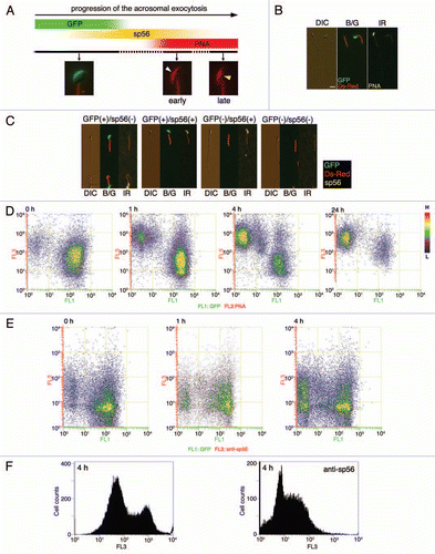
Figure 2 Bead-MIVF assay. (A) Low magnification views (left and center) of the COC in medium containing protein-coupled fluorescent beads. Images were taken by differential interference contrast microscopy (DIC) and fluorescence microscopy with an ultraviolet (UV) filter. An outer edge of the CCL (center, inset) was enlarged (right). No beads were found in the CCL, indicating that 0.5 µm polystyrene microparticles (Polysciences) cannot penetrate the cumulus matrix by free diffusion. (B) Capacitated spermatozoa were thereafter inseminated and their entry into the CCL was monitored to determine if the beads were carried by CCL-entering sperm. Beads used were control IgG beads, anti-sp56 beads and PNA beads. Photographs were taken every 5 min using fluorescence microscopy with a UV filter or a dual band-pass filter (B/G). (C) After 30 min of insemination, the fluorescent beads and sperm within the CCL were counted and the bead/sperm ratio calculated. The data are expressed as the mean ratio of beads/sperm ± SEM obtained from 4 independent experiments. Scale bars: 50 µm.
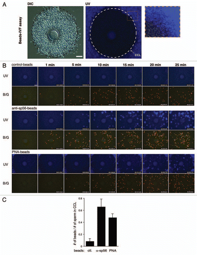
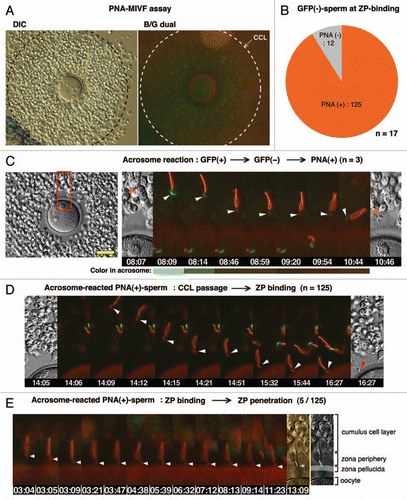
Figure 3 PNA-MIVF assay. (A) Low magnification views of the COC in medium containing Alexa 594-labeled PNA under DIC (left) and fluorescence (right) microscopy with a dual band-pass filter (B/G). An outer border of the CCL was marked (dashed circle). In contrast to the bead-IVF assay, PNA could penetrate the cumulus matrix and stained the ZP. In comparison to the high red fluorescence background (due to presence of Alexa-PNA) in the CCL, the zona periphery was relatively dark, providing an opportunity to detect the acrosomal status of sperm arriving at this area. (B) Among EGFP− sperm that bound to the ZP, most were PNA+, whereas approximately 10% were PNA−. (C) A representative case of a sperm that underwent the AR while traveling through the zona periphery. Shown are the full screen image of the COC in this experiment (left) and enlarged (boxed area) time-lapse images of the spermatozoon undergoing the AR (right). A gradual disappearance of the acrosomal green fluorescence (8 min 9 s) followed by accumulation of red fluorescence (9 min 54 s) was observed. Color in the acrosome was shown as a bar below each photograph. (D) Representative photographs of the consecutive processes from sperm passage through the CCL to sperm binding to the ZP by PNA+ sperm. (E) Out of 125 PNA+, ZP-bound sperm, 5 sperm initiated ZP-penetration. Arrowheads point to the acrosomal region; scale bar, 50 µm.
Addendum to: