
Abstract
GSH is mostly considered a non-enzymatic antioxidant that serves for modulating the redox status of protein thiols, detoxification, and direct scavenging activity of oxyradicals. Within the cells, GSH has also the role to buffer the flux of nitric oxide (NO), which in the nervous system is physiologically produced being an important neuromodulator and neurotransmitter. However, this role of GSH in modulating NO toxicity is often considered of secondary importance. Recently, we confuted such assumption as we demonstrated that GSH depletion triggers a severe NO imbalance, which is the primary cause of neuronal death. Here we report that even a slight and non-toxic decrease of GSH in brain mice causes protein nitration that is reversed by inhibiting NO production. This evidence indicates that NO imbalance and the associated nitrosative hallmarks observed in neurodegenerative diseases as well as in health ageing are likely the consequence of the progressive decline of GSH.
Acknowledgments
This work was partially funded by grants from Ministero della Salute (#GR-2008-1138121) and MIUR.
Figures and Tables
Figure 1 Glutathione depletion is associated with 3-nitrotyrosine increase in mice brain without accumulation of apoptotic markers. (A) Left part: 20 µg of proteins extracted from mice brains were spotted on nitrocellulose membrane and subjected to Dot blot analysis using a polyclonal 3-nitrotyrosine (3-NT) antibody. Right part: density of immunoreactive dots was normalized for Ponceau Red spots and reported as arbitrary units (a.u.) and as means ± SD. (*p < 0.01 versus control, **p < 0.01 versus BSO-treated; n = 3). (B) 20 µg of total protein extracts were loaded for detection of Bcl-2, Bax and pro-caspase-3 by western blot. Actin was used as loading control. (C) mitochondria were purified from mice brains homogenates by Percoll® gradient, lysed and 20 µg of proteins were loaded for detection of AIF by western blot. Cytochrome c was used as loading control.
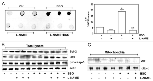
Table 1 GSH concentration in mice treated with BSO
Addendum to: