
Abstract
Despite a long history of anatomical mapping of neuronal networks, we are only beginning to understand the detailed three-dimensional (3D) organization of the cortical micro-circuitry. This is in part due to the lack of complete reconstructions of individual cortical neurons. Morphological studies are either performed on incomplete cells in vitro, or when performed in vivo, lack the necessary cellular resolution. We recently reconstructed the in vivo axonal and dendritic morphology of two types of L(ayer) 5 neurons from vibrissal cortex. The 3D profiles of short-range as well as long-range projections indicate that L5 slender-tufted and L5 thick-tufted neurons represent very different building blocks of the cortical circuitry. In this addendum to Oberlaender et al, PNAS 2011, we motivate our technical approach and the advancements this may give in reconstructing the cortical micro-circuitry.
Acknowledgments
This work was supported by the Max-Planck Society, the Center for Neurogenomics and Cognitive Research at Vrije Universiteit Amsterdam, and a Veni Grant from The Netherlands Organization for Scientific Research (to C.P.J.d.K.).
Figures and Tables
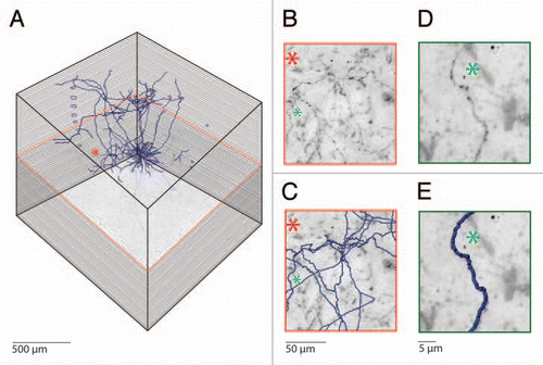
Figure 1 Semi-automated reconstruction of a Layer 5 pyramidal neuron filled with biocytin in vivo. (A) Mosaic scanning and subsequent serial reconstruction is performed on consecutive 100 µm thick tangential sections, which results in high resolution 3D images representing cubic millimeters of cortical volumes. The consecutive slices are aligned by using blood vessels that run perpendicular to the cortical surface. (B) Magnification of the area indicated by the asterisk. Note the abundant axons running through this area. (C) Automated detection of biocytin labeled processes allows fast and reliable reconstruction of axonal morphology. Axonal reconstructions from additional Z values are also visible in this part. (D) Example images of a single axonal branch labeled with biocytin and (E) the subsequent automated reconstruction.
Addendum to: