Abstract
Chronic pain is characterized by post-injury pain hypersensitivity. Current evidence suggests that it might result from altered neuronal excitability and/or synaptic functions in pain-related pathways and brain areas, an effect known as central sensitization. Increased activity of extracellular signal-regulated kinase (ERK) has been well-demonstrated in the dorsal horn of the spinal cord in chronic pain animal models. Recently, increased ERK activity has also been identified in two supraspinal areas, the central amygdala and the paraventricular thalamic nucleus anterior. Our recent work on the capsular central amygdala has shown that this increased ERK activity can enhance synaptic transmission, which might account for central sensitization and behavior hypersensitivity in animals receiving noxious stimuli.
Figures and Tables
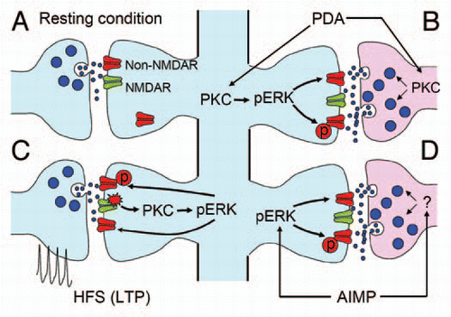
Figure 1 Schematic diagram summarizing PBA-CeAC synaptic transmission in normal and AIMP mice. (A) Synaptic transmission in a resting condition at PBA-CeAC synapses is mediated by non-NMDARs. (B) Application of PDA activates PKC in presynaptic terminals and postsynaptic dendrites. Activated PKC in presynaptic terminals enhances glutamate release, while that activated in postsynaptic spines activates ERK to upregulate non-NMDAR functions or increase their numbers at synaptic sites. (C) High-frequency stimulation activates NMDARs, which in turn activate the PKC-ERK signal pathway to upregulate non-NMDAR functions or increase their numbers at synaptic sites, thereby resulting in LTP. (D) In mice with AIMP, ERK is activated in the CeAC by an excessive nociceptive signal, which in turn upregulates non-NMDAR functions or increases their numbers at synaptic sites. In addition, enhanced glutamate release from presynaptic terminals was also observed in AIMP mice. Together, these pre- and postsynaptic enhancements at PBA-CeAC synapses might partially account for the central sensitization in AIMP.