
Abstract
Measurement of the activity of neuronal ensembles is an essential step to understand how the neuronal network is organized and functioning. Electrical excitation of neurons causes calcium influx via voltage-gated calcium ion channels, which can be monitored by calcium imaging using fluorescent calcium probes. DNA-encoded calcium indicators (DECIs) such as cameleon and GCaMP have been developed to specifically label a subpopulation of neurons. However, in many cases, DECIs that had been developed and tested in vitro did not always show expected performance in vivo. It is necessary to increase its sensitivity and also to adjust its dynamic range to the physiological conditions. In our recent study, we developed an improved version of GCaMP and tested its performance in vivo using transgenic zebrafish. By combining the new GCaMP with targeted gene expression via the Gal4FF-UAS system, we successfully imaged the activity of the spinal motor circuit during spontaneous contractions of zebrafish larvae. Further we report here that heptanol, a gap junction blocker, could alter the spatiotemporal activation pattern of the motor circuit. Thus, we demonstrate that calcium imaging with GCaMP is powerful to analyze neuronal activities under normal and pharmacologically perturbed conditions.
Acknowledgements
We thank A. Ito, Y. Kanebako, N. Mouri, M. Mizushina and M. Suzuki for fish room works, H. Takakubo for technical assistance, the Mitsubishi Foundation (to A.M.) and the National BioResource Project and grants from the Ministry of Education, Culture, Sports, Science and Technology of Japan.
Figures and Tables
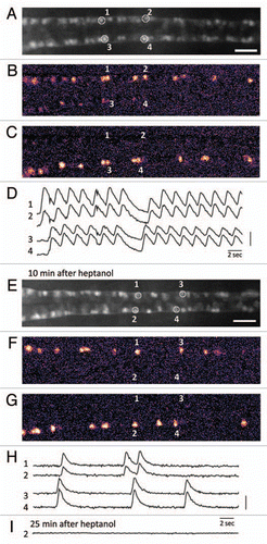
Figure 1 Calcium imaging of the motor circuit in the zebrafish spinal cord during spontaneous contractions. (A) Expression of GCaMP-HS in the CaP motor neurons in the spinal cord. A dorsal view of the SAIG213A;UA S:GCaMPHS double transgenic embryo embedded in 2% low-melting agarose under a fluorescence microscope and treated with a neuromuscular junction blocker, D-tubocurarine. CaP neurons #1–#4 were used as ROIs (regions of interest). Anterior to the left. Scale bar: 200 µm. (B and C) Calcium signals of the CaP motor neurons with pseudocolors (see also www.youtube.com/watch?v=6y44uxrh7z4). (B) The CaP motor neurons on the right side including ROI-1 and -2 showed increased fluorescence. (C) The CaP motor neurons on the left side including ROI-3 and -4 showed increased fluorescence. (D) The fluorescence changes in the selected CaP motor neurons. 1 and 2 (3 and 4) are activated synchronously, and the right (1 and 2) and left (3 and 4) neurons are activated alternately. A vertical bar indicates (F-resting F)/resting F = 50%. (E) A dorsal view of the SAIG213A;UA S:GCaMPHS double transgenic embryo embedded in 2% low-melting agarose under a fluorescence microscope and treated with a gap junction blocker, heptanol, for 10 min. Anterior to the left. Scale bar: 200 µm. (F and G) Calcium imaging of the CaP motor neurons in the presence of heptanol with pseudocolors (see also www.youtube.com/watch?v=3xhw9D35H5w). (F) The CaP motor neurons on the right side including ROI-1 and -2 showed increased fluorescence. (G) The CaP motor neurons on the left side including ROI-3 and -4 showed increased fluorescence. (H) The fluorescence changes in the selected CaP motor neurons. Synchronized activation is still observed. A vertical bar indicates (F-resting F)/resting F = 100%. (I) The fluorescence changes are not detected 25 min after the heptanol treatment.
Addendum to: