
Abstract
We propose a new in vitro system to study the mechanics of surface area regulation in cells, which takes into an account the spatial confinement of the cell membrane. By coupling a lipid bilayer to the strain-controlled deformation of an elastic sheet, we show that upon straining the supported lipid bilayer expands its surface area by absorbing adherent lipid vesicles and upon compression decreases its area by expelling lipid tubes out of its plane. The processes are reversible and closely resemble in vivo observations on shrinking cells. Our results suggest that the mechanics of the area regulation in cells is controlled primarily by the membrane tension and the effects of the membrane confinement.
Acknowledgments
We thank M. Arroyo, D.P. Holmes and C. Read for discussions and Princeton University and the Project X Fund for financial support.
Figures and Tables
Figure 1 Schematic of the absorption of a giant vesicle (green) into laterally stretched supported bilayer (red). The process passes through the stages (i) adhesion, (ii) hemifusion of the membranes in the contacting area (yellow zones), (iii) pore formation on the unadhered portion of the vesicle membrane and consequent content leakage, (iv) collapse of the vesicle and (vi) final absorption, and as such differs from the commonly observed fusion mechanism through the formation of a fusion pore (inset).
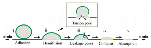
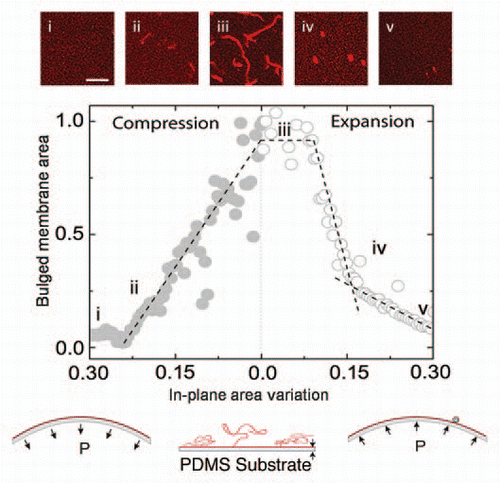
Figure 2 Dynamic transformation of a supported lipid membrane throughout a cycle of area compression and expansion (schematic insets). The area of the out of plane lipid tubes during elongation (compression) and retraction (expansion) is shown as a function of the in-plane membrane area. The insets are confocal images of (i) the bilayer in the initial unstrained conditions, (ii) tube nucleation at a critical compressive threshold, (iii) elongated tubes as a result of the membrane compression, (iv) retraction of tubes to vesicles under membrane expansion and (v) further absorption of the vesicles into the expanding bilayer. Scale bar: 20 microns.
Addendum to: