
Abstract
Histone deacetylases (HDACs) are enzymes that catalyse the removal of acetyl groups from a range of nuclear and cytoplasmic proteins. Recently, we described a novel route to neurotrophin-dependent gene activation in neurons, which requires the S-nitrosylation of nuclear HDAC2 by the gaseous molecule nitric oxide (NO) (Nott et al, 2008). We have further investigated the NO-dependent regulation of HDACs in neurons. Using a fluorogenic deacetylation assay, we show that NO decreases the enzymatic activity of a sub-group of neuronal HDACs in vitro and that this inhibition is not due to damaging modifications such as oxidation or tyrosine nitration. The neuronal HDACs whose catalytic activity is inhibited by NO are entirely those that are localised in the cytoplasm. These observations support and extend the concept that nitric oxide is a key regulator of HDAC function in mammalian neurons.
Acknowledgements
We would like to thank Andrew Porter for assistance in carrying out experiments analyzing the effects of nitric oxide on tubulin acetylation. This work was supported by the Medical Research Council (Research Grant G0500792) and the European Research Council (Marie Curie International Reintegration Grant MIRG-CT-2005-016501). P. Marc D. Watson is the recipient of a Medical Research Council Career Development Fellowship. Antonella Riccio is a recipient of an MRC Career Development Award (G117/533).
Figures and Tables
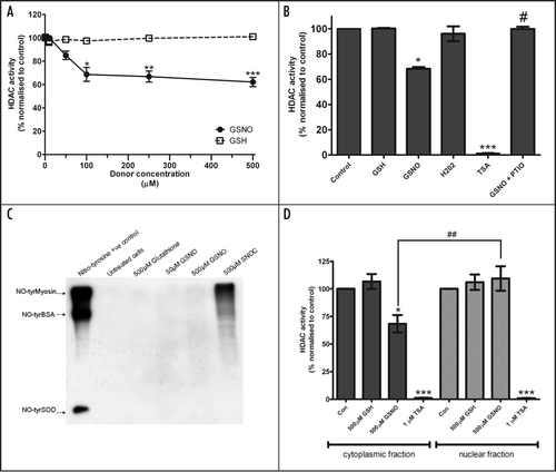
Figure 1 (A) Whole cell lysates prepared from E18 embryonic cortical neurons were used in a fluorogenic HDAC assay as previously shown.Citation1,Citation26 Lysates were incubated with the NO donor GSNO, or control glutathione (GSH) for 1 hour at room temperature before HDAC activity was assayed. HDAC activity was normalised to untreated control (100% activity). Data were analysed using paired t-tests comparing GNSO vs. GSH-treated lysate at each concentration point (n = 5). *p = 0.0161; **p = 0.0085; ***p = 0.0006. (B) Whole cell neuronal lysates were incubated with GSNO, GSH, hydrogen peroxide(all 500 µM), TSA (1 µM) and GSNO (500 µM) in the presence of the NO scavenger PTIO (200 µM) for one hour at room temperature. Data are normalized to untreated (100%) and presented as mean ± SEM (n = 3): *p < 0.05 and ***p < 0.001 (versus control), #p < 0.05 (GSNO vs. GSNO + PTIO) by one-way ANOVA with Tukey's post hoc test. (C) Anti-nitrotyrosine western blot analysis of neuronal lysates following 1 hour incubation with NO donors. Tyrosine-nitrated myosin, BSA and superoxide dismutase were used as positive control. Tyrosine nitration was not observed with concentrations of GSNO up to 500 µM (n = 2). Some tyrosine nitration appeared following treatment with S-nitrosocysteine (SNOC). (D) Neuronal cytoplasmic and nuclear extracts were incubated with GSNO (500 µM), GSH (500 µM) or TSA (1 µM) for 1 hour at room temperature and subjected to fluorogenic HDAC assay. Data are presented as mean ± SEM (n = 3) after normalisation to untreated (100%) HDAC activity. Statistical differences were calculated using one-way ANOVA with Tukey's post hoc test: *p < 0.05 and ***p < 0.001 (versus control), ##p < 0.01 (500 µM GSNO vs. 500 µM GSH).
Addendum to: