
Abstract
Neuronal adaptation has been studied extensively in visual motion-sensitive neurons of the fly Calliphora vicina, a model system in which the computational principles of visual motion processing are amenable on a single-cell level. Evidenced by several recent papers, the original idea had to be dismissed that motion adaptation adjusts velocity coding to the current stimulus range by a simple parameter change in the motion detection scheme. In contrast, linear encoding of velocity modulations and total information rates might even go down in the course of adaptation. Thus it seems that rather than improving absolute velocity encoding motion adaptation might bring forward an efficient extraction of those features in the visual input signal that are most relevant for visually guided course control and obstacle avoidance.
Acknowledgements
Thanks to C. Spalthoff for his contribution to the illustration.
Figures and Tables
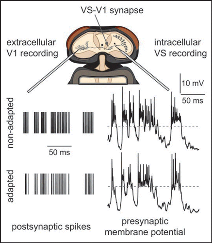
Figure 1 Adaptation of fly visual motion-sensitive neurons. Two synaptically coupled types of neurons were simultaneously recorded in vivo during presentation of a grating pattern. Repeated identical sequences of random velocity modulations of the pattern were presented. Top, schematic of the fly head with the exposed recording site. Bottom, membrane potential responses of the presynaptic VS-neuron (right) and trains of action potentials of the postsynaptic V1-neuron (left). VS-neurons transfer signals at synapses by graded potentials. Dashed horizontal line represents resting membrane potential of VS prior to stimulation. Action potentials of V1 are indicated by vertical lines.
Addendum to: