Abstract
Comparative molecular, developmental, and morphogenetic analyses show that the three major segmented animal groups – Lophotrochozoa, Ecdysozoa, and Vertebrata - use a wide range of ontogenetic pathways to establish metameric body organization. Even in the life history of a single specimen, different mechanisms may act on the level of gene expression, cell proliferation, tissue differentiation, and organ system formation in individual segments. Accordingly, in some polychaete annelids the first three pairs of segmental peripheral neurons arise synchronously, while the metameric commissures of the ventral nervous system form in anterior-posterior progression. Contrary to traditional belief, loss of segmentation may have occurred more often than commonly assumed, as exemplified in the sipunculans, which show remnants of segmentation in larval stages but are unsegmented as adults. The developmental plasticity and potential evolutionary lability of segmentation nourishes the controversy of a segmented bilaterian ancestor versus multiple independent evolution of segmentation in respective metazoan lineages.
Acknowledgements
Research in the lab of Andreas Wanninger is funded by the EU Early Stage Research Training Network MOLMORPH under the 6th Framework Programme (contract number MEST-CT-2005—020542). Both Alen Kristof and Nora Brinkmann are recipients of a fellowship within the MOLMORPH programme.
Figures and Tables
Figure 1 Schematic representation of neurogenesis in the polychaete annelid Sabellaria alveolata based on serotonin immunoreactivity, revealing differences in the mode of establishment of metamery in the peripheral segmental neurons and the ventral commissures, respectively. Both aspects are ventral views with anterior facing upwards. Total length of the specimens is approximately 280 µm in (A) and 330 µm in (B). (A) Late larva with synchronously established peripheral segmental neurons (yellow). Ventral commissures and perikarya along the paired ventral nerve cord (vnc) are still lacking. The prototroch nerve ring (pnr) and the nerve ring underlying the telotroch (ttn) constitute subsets of the larval nervous system, while the circumoesophageal commissures (cc) and the longitudinal trunk neurons (ltn) are parts of the adult neural bodyplan. (B) Larva prior to metamorphosis. The ventral commissures (asterisks) of the first five segments have been established progressively, together with the paired, metameric sets of perikarya (red dots) along the ventral nerve cords (vnc). The six pairs of peripheral segmental neurons (yellow) correspond to the segments II–VII, because development of segment I is retarded in this species, resulting in development of the paired peripheral segmental neuron of this segment at a later stage. Note that ontogeny of the peripheral segmental neurons precedes development of the ventral commissures in segments VI and VII. pns - the nerves of the peripheral nervous system.
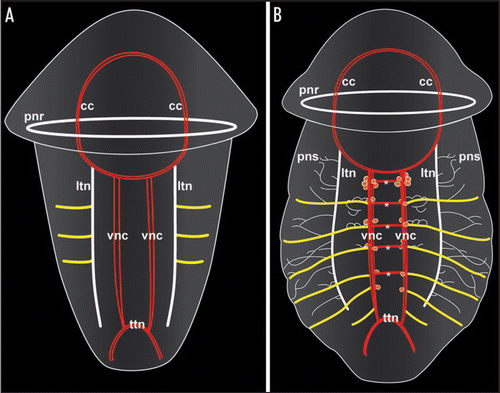
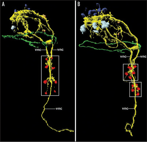
Figure 2 Expression and loss of the segmental pattern of the ventral CNS in the sipunculan Phascolosoma agassizii, as depicted by 3D reconstruction of serotonin immunoreactivity. Both aspects are ventral views with anterior facing upwards. Total length of the specimens is approximately 150 µm. (A) Late larva with four pairs of metameric perikarya (red; boxed area) associated with the ventral nerve cord (vnc). The latter is already fused along the entire anterior-posterior axis except in the anterior-most region. Additional neural elements include the cells of the larval apical organ (dark blue) overlying the neuropil mass (np) of the adult brain, the first cell bodies of the developing adult brain (light blue), two cells of the peripheral nervous system (orange), and the larval prototroch nerve ring (green). (B) Larva prior to metamorphosis in which the metameric arrangement of the ventral perikarya (red) has been lost in favor of two cell clusters (boxed areas) comprising five cells each. Note the increased number of cells belonging to the adult brain (light blue).