
Abstract
The plasma membrane of mammalian cells is composed of a great variety of different lipids which are laterally organized into lipid domains. The segregation of lipids into domains has been studied in great detail in vesicles but domain formation of lipids in the plasma membrane of live cells is still unclear. We have previously used fluorescence lifetime imaging microscopy to study the colocalization of the receptor for EGF with the ganglioside GM1 and the GPI-anchored green fluorescent protein. Here we have used this technology to study the effect of EGF on the organization of GM1 in the plasma membrane. Our data show that stimulation of the cell with EGF induces rapidly a strong increase in colocalization of GM1 molecules, suggesting the formation of large lipid domains. These results support the notion that activation of EGFR signaling may result in the formation of signaling platforms.
Acknowledgements
This work was supported by the ‘From Molecule to Cell’ program from the Dutch scientific organisation (NWO-ALW, grant 805.47.084) to Erik Hofman and Arjen Bader.
Figures and Tables
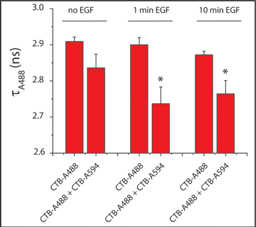
Figure 1 EGF stimulation induces coalescence of GM1-containing rafts. Serum-starved HER14 cells (NIH 3T3 cells stably expressing human EGFR), were labeled with 1 µg/ml cholera toxin B-subunit conjugated to Alexa Fluor 488 (CTB-A488) and/or Alexa Fluor 594 (CTB-A594) on ice. After 1 hour, the cells were recovered to 37°C and stimulated with 20 ng/ml EGF for 1 or 10 minutes. The cells were fixed with 4% formaldehyde and the coverslips were embedded in mowiol. The lifetimes of Alexa 488 were determined by FLIM analysis as described previously.Citation9 Histograms show the average lifetime values of four cells per condition (*p < 0.05).
Addendum to: