
Abstract
The two fundamental lineages of photoreceptor cells, microvillar and ciliary, were long thought to be a prerogative of invertebrate and vertebrate organisms, respectively. However evidence of their ancient origin, preceding the divergence of these two branches of metazoa, suggests instead that they should be ubiquitously distributed. Melanopsin-expressing ‘circadian’ light receptors may represent the remnants of the microvillar photoreceptors amongst vertebrates, but they lack the characteristic architecture of this lineage, and much remains to be clarified about their signaling mechanisms. Hesse and Joseph cells of the neural tube of amphioxus (Branchiostoma fl.) – the most basal chordate extant - turn out to be depolarizing primary microvillar photoreceptors, that generate a melanopsin-initiated, PLC-dependent response to light, mobilizing internal Ca and increasing a membrane conductance selective to Na and Ca ions. As such, they represent a canonical instance of invertebrate-like visual cells in the chordate phylum.
Figures and Tables
Figure 1 Simplified phylogenetic tree. Ciliary photoreceptors are typical of vertebrata (in the chordata phylum), whereas microvillar photoreceptors have been extensively characterized both morphologically and physiologically in arthropoda and mollusca. However, putative photoreceptors of both types have subsequently been identified also in pre-bilateria, such as box jellyfish (cnidaria).
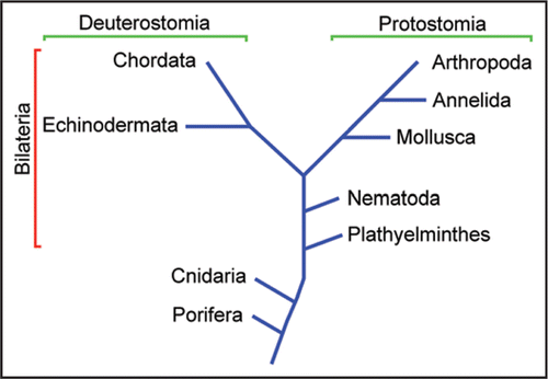
Figure 2 The amphioxus (Branchiostoma floridae). (A) Intact specimen. Calibration bar: 5 mm (B) Joseph cell enzymatically dissociated from the neural tube (the shadow is a recording patch microelectrode). (C) Isolated organ of Hesse, comprised of a pigmented cell and a separate, microvilli-bearing translucent cell. Calibration bars in (B and C): 10 µm.
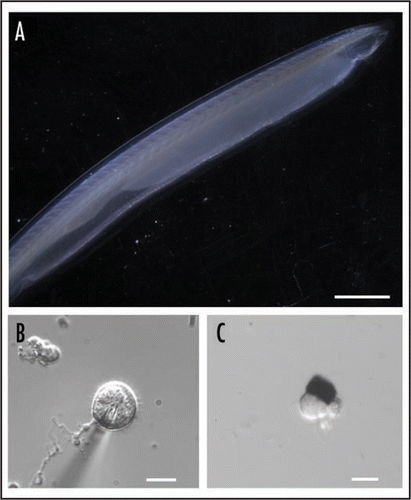
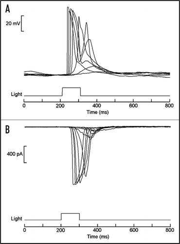
Figure 3 Light responses in isolated organ of Hesse. (A) Superimposed traces of membrane voltage recording, showing depolarization elicited by brief flashes of light. (B) Light-activated inward currents measured under voltage clamp by the whole-cell patch recording technique. In both cases stimuli were delivered every minute, and the intensity of the light was increased at 0.6 log increments. Similar responses were also obtained from Joseph cells.
Addendum to: