
Abstract
Myosin-1a is one of eight monomeric, membrane binding class I myosins expressed in vertebrates.1 As the most abundant actin-based motor protein found in the enterocyte microvillus, myosin-1a has long been known to interact with the apical membrane via a highly basic C-terminal tail domain.2 Several recent studies shed light on possible functional consequences of this protein/lipid interaction. In vitro and in vivo studies of microvillar function have revealed that myosin-1a can move apical membrane along core actin bundles, leading to the release of small vesicles from microvillar tips.3,4 Additional studies indicate that myosin-1a and other class I myosins contribute to membrane-cytoskeleton adhesion, which enables the apical membrane to resist deformation.5 These findings clearly position myosin-1a as an important player in apical membrane movement and structural stability. How this motor is able to fulfill these two seemingly distinct functions is currently unclear, but will serve as the focus of our discussion below.
Acknowledgements
This work was supported by grants from the National Institutes of Health (R01 DK-075555, M.J.T.) and the American Heart Association (09GRNT2310188, M.J.T.; Post-doctoral Fellowship 0825358E, R.N.).
Figures and Tables
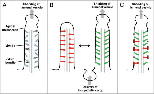
Figure 1 Models of Myo1a multifunctionality. (A) Single population of asynchronously cycling/functioning Myo1a molecules. In this model, all myosin molecules are functionally equivalent, participating in both the tip-ward movement of apical membrane and M-C adhesion while strongly bound to actin (molecules with black outline). Asynchronous AT Pase cycling means that a fraction of the total Myo1a population will always be strongly bound to the actin core bundle. The strongly bound fraction is controlled by the Myo1a duty ratio, which has the potential to increase dramatically if interactions with the apical membrane present even a minor opposing load. (B) Single Myo1a population switching between two functional states. Red Myo1a molecules are those contributing to M-C adhesion, green molecules are driving tip-ward movement of apical membrane. Delivery of biosynthetic vesicles to the inter-microvillar region transiently reduces tension in the apical membrane and impedance to Myo1a cycling; this gives rise to membrane movement, which leads to the formation of a vesicle at the microvillus tip. (C) Coexistence of two functionally distinct Myo1a populations. The thicker red membrane regions represent detergent insoluble patches that may immobilize a subset of Myo1a molecules (red), allowing them to contribute to M-C adhesion. Myo1a not bound to these patches would be more dynamic (green molecules) and contribute to the tip-ward flow of apical membrane components.
Addendum to: