
Abstract
Presynaptic N-type voltage-gated Ca2+ channels (CaV2.2) form part of an extensive macromolecular complex in the presynaptic terminal. Regulation of CaV2.2 is achieved via protein-protein interactions within the terminal and can directly impact transmitter release which is dependent on Ca2+ influx via these CaV2.2. We recently identified a novel CaV2.2 interacting partner – the collapsin response mediator protein (CRMP).1 CRMPs are a family of five proteins implicated in signal transduction of neurite outgrowth and axonal guidance. We showed that CRMP-2, a well-studied member of this family, interacted with CaV2.2 via direct binding to cytoplasmic loops of CaV2.2. Depolarization enhanced the interaction. Further studies revealed that CRMP-2 facilitated an increase in CaV2.2 current density by inserting more CaV2.2 at the cell surface. As a consequence of CRMP-2-mediated increase in Ca2+ influx, release of the excitatory neurotransmitter glutamate was also increased. CRMP-2 localized to synapses where, surprisingly, its overexpression increased synapse size. We hypothesize that the CRMP-2–calcium channel interaction represents a novel mechanism for modulation of Ca2+ influx into nerve terminals and, hence, of synaptic strength. In this addendum, we further discuss the significance of this study and the possible implications to the field.
Acknowledgements
CRMP4a cDNA was a gift of Dr. Alyson Fournier (Montreal Neurological Institute, Montreal, Canada). We thank Drs. Gerry Oxford and May Khanna and Lisa D. King and Brian W. Jarecki for critically reading the manuscript. This work was supported by grants from the Indiana State Department of Health—Spinal Cord and Brain Injury Fund [Grant 4786219 to R.K.] and The Indiana University Biomedical Committee—Research Support Funds [Grant 2286501 to R.K.].
Figures and Tables
Figure 1 CRMP-2 signaling cascade: a novel role for CRMPs in Ca2+ channel regulation and transmitter release. Extracellular signals, such as extracellular matrix, growth factors and guidance cues (semaphorin 3A) activate Neuropilin-1/Plexin A receptors on membranes.Citation7 A battery of kinases, including RhoK, Cdk5 and GSK-3β phosphorylate CRMPs. Phosphorylated CRMPs have a reduced affinity to tubulin and other interacting molecules and lose their positive effect on axon elongation, thereby causing growth arrest and growth cone collapse. In contrast, non-phosphorylated CRMPs bind strongly to tubulin heterodimers to promote microtubule assembly and Numb-mediated endocytosisCitation30 thereby promoting axon elongation and branching.Citation7 In addition to these classically defined roles for CRMPs, our results suggest that CRMPs (assuming both phosphorylated and non-phosphorylated forms) bind to cytoplasmic loops of the Ca2+ channel and increase their insertion into the membrane, resulting in an increased current density.Citation1 This increase culminates into an increase in the release of the excitatory transmitter glutamate.Citation1 Interestingly, CRMP-2 overexpression increases synapse size not number.Citation1 This suggests that CRMP-2 regulation of transmitters may occur via a direct effect on CaV2.2 or through an effect on changes in synaptic vesicle machinery and release probabilities. Increased synaptic transmission is likely to contribute to synaptic plasticity.
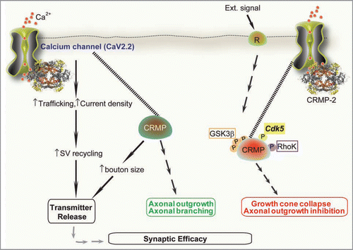
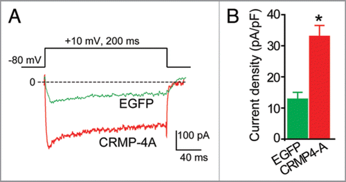
Figure 2 CRMP-4 enhances Ca2+ current density in hippocampal neurons. (A) Exemplar current traces obtained from a cell transfected with EGFP and CRMP-4-EGFP evoked by 200-ms steps to +10 mV applied from a holding potential of -80 mV, as shown in the voltage protocol above the traces. Bath solutions contained 1 µM TT X, 10 mM TE A and 1 µM Nifidepine to block Na+, K+ and L-type voltage-gated Ca2+ channels, respectively. (B) Peak current density (pA/pF) measured at +10 mV for EGFP (n = 8) or CRMP-4a-EGFP (n = 8) transfected neurons. *p < 0.05 versus EGFP (One-way ANOVA).
Addendum to: