Abstract
Exosomes are endosome-derived vesicles (40-100nm) formed during the formation of multi-vesicular bodies (MVBs). Occasionally, the MVBs fuse with the plasma membrane releasing their intra-luminal vesicles into the extracellular media, which are then known as exosomes. Different cell types such as B-cells, dendritic cells, platelets, reticulocytes and macrophages can release exosomes and current research in this area is more focused towards exosomes released by antigen-presenting cells. Exosomes have recently been shown to be immunomodulatory and the mechanism of immune response initiation by them is beginning to emerge. Besides molecules present inside the lumen of exosomes, it has been suggested that certain exosomal membrane molecules can interact with their surface receptors on the target cells thereby inducing an immunomodulatory response. In this review, Hsp70 and galectin-5, two immunogenic molecules present on exosomal membrane, are discussed in detail for initiating this response.
Acknowledgements
The author would like to thank Gareth Griffiths (Institute for Molecular Biosciences, Oslo, Norway) for helpful discussions; David Liebl (Institute for Molecular Bioscience, Brisbane, Australia) and A.A. Jeyaprakash (Max-Planck Institute, Martinsried, Germany) for critical reading of the manuscript, and Christopher K.E. Bleck (C-CINA, University of Basel, Basel, Switzerland) for providing electron micrograph in . This work was supported by Alexander von Humboldt Foundation, Germany and European Union Sixth Framework Program.
Figures and Tables
Figure 1 EM image showing presence of Hsp70 on exosome membrane. RAW 264.7 mouse macrophages were given heat-shock at 43°C for 2 h. After 6 h recovery at 37°C, exosomes were purified from culture supernatants by differential centrifugation and floatation on a sucrose gradient. The samples were processed for surface immunogold labeling with anti-Hsp70 antibody. Arrows show Hsp70 (gold) labeling on exosome surface. Insets show magnified images of exosomes with surface Hsp70 labeling. (Micrograph provided by Christopher K.E. Bleck).
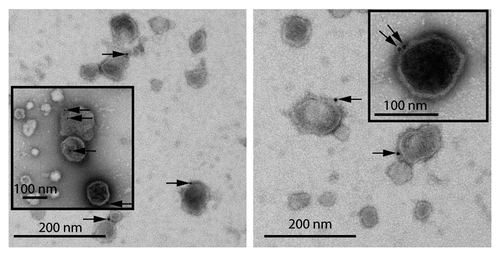
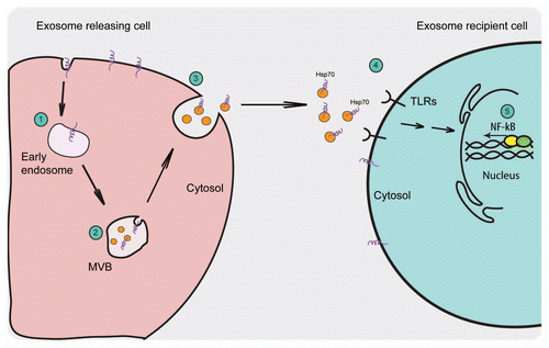
Figure 2 Schematic representation of the exosome release pathway and the proposed activation of the immune response by exosomal Hsp70. (1) Extracellular molecules or membrane proteins are internalized into the endosomes that form intra-luminal vesicles by the inward budding of their limiting membrane. (2) Under certain conditions, these MVBs fuse with the plasma membrane releasing their ILVs to the outside where they are known as exosomes. (3) Exosomes exhibit the same orientation of their membrane proteins as on the plasma membrane with extracellular domain of the protein present on the exosomal surface. (4) Exosomes that are Hsp70-positive can interact with TLRs on the cell surface (5) thus activating NF-⊠B signaling pathway.