
Abstract
Defective autophagy and lysosomal degradation are hallmarks of numerous neurodegenerative disorders. Vesicular ATPases are intracellular proton pumps that acidify autophagosomes and lysosomes. V0a1 is a key component of the v-ATPase that is only required in neurons in Drosophila melanogaster. We have recently shown that loss of V0a1 in Drosophila photoreceptor neurons leads to slow, adult-onset degeneration.1 Concurrently, Lee et al.2 reported that V0a1 fails to localize to lysosomal compartments in cells from Presenilin 1 knock-out cells. Together these two reports suggest that a neuronal V0a1-dependent degradation mechanism may be causally linked to Alzheimer pathology. Indeed, we now show that loss of V0a1 makes Drosophila neurons more susceptible to insult with human Alzheimer-related neurotoxic Aβ and tau proteins. Furthermore, we discuss the potential significance of the discovery of the neuron-specific degradation mechanism in Drosophila for intracellular degradation defects in Alzheimer Disease.
Acknowledgements
We would like to thank Drs. Helmut Kramer, Damian Crowther, Craig Montell, the Bloomington Stock Center, and the University of Iowa Developmental Studies Hybridoma Bank for reagents. We further thank Drs. Ilya Bezprozvanny and Adam Haberman for comments on the manuscript and discussion. This work was supported by grants from the National Institute of Health (RO1EY018884) and the Welch Foundation (I-1657). P.R.H. is a Eugene McDermott Scholar in Biomedical Research.
Figures and Tables
Figure 1 Loss of the v0a1-dependent degradation pathway increases susceptibility to neurotoxic insults by human Aβ and tau proteins. (A) Representative ERG recording for at least n = 20 three day-old flies of the genotypes indicated and raised at room temperature under constant light stimulation. (B) Quantification of ERG depolarizations. Orange bar shows ERG depolarization for control or overexpressed proteins in wild type photoreceptors; blue bars are corresponding experiments with dicer2-enhanced v0a1 rNAi. Asterisks show statistical significance in pairwise comparisons (p < 0.005). (C) Immunolabeled adult eyes from experimental flies used for ERG recordings shown in (A). Chaoptin in green and nuclear labeling with Toto-3 in blue; Syx7/Avl labeling in red. Arrows indicate degenerative ‘holes’ in the immunolabeling. Arrowheads indicate early endosomal Syx7-positive accumulations with Chaoptin that are typical for loss of v0a1 function. Error bars are S.E.M.
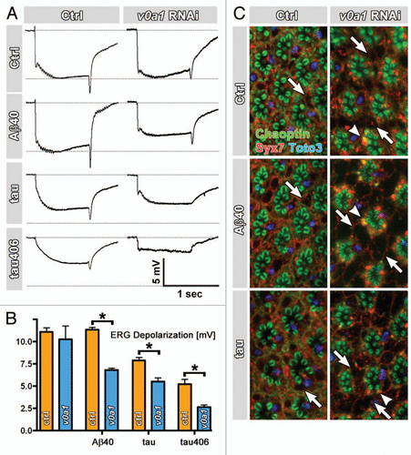
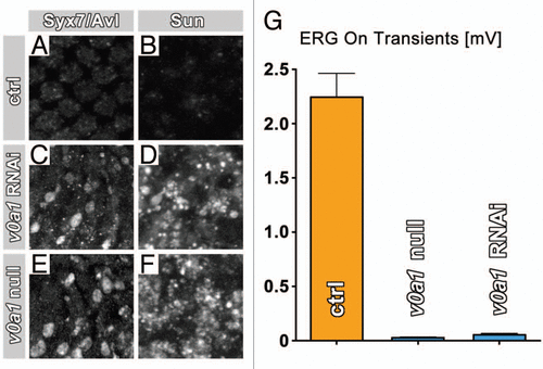
Figure 2 v0a1 rNAi in photoreceptors causes phenotypes identical to loss of v0a1. (A–F) Antibody-labeling of Syx7/Avl and Sunglasses reveals similar accumulations of both markers in photoreceptors expressing dicer2-enhanced v0a1 RNAi (C and D) as in v0a1null mutant photoreceptors in ey35FLP v0a1 flies (E and F). (G) Both v0a1 RNAi as well as ey35FLP v0a1 lead to a complete loss of neurotransmission, as indicated by the loss of electroretinogram (ERG) ‘on’ transients.
Addendum to: