
Abstract
Specialized neuronal structures namely growth cones, filopodia and spines are important entities by which neurons communicate with each other, integrate multiple signaling events, consolidate interacting structures and exchange synaptic information. Recent studies confirmed that Transient Receptor Potential Vanilloid sub type 1 (TRPV1), alternatively known as capsaicin receptor forms a signaling complex at the plasma membrane and integrate multiple exogenous and endogenous signaling cues there. This receptor localizes in the neuronal growth cones and also in filopodial tips. In addition, TRPV1 is endogenously present in synaptic structures and located both in pre- and post-synaptic spines of cortical neurons. Being non-selective Ca2+-channel, TRPV1 regulates the morphology and the functions of these structures by various mechanisms. Our studies indicated that physical interaction with signaling and structural molecules, modulation of different cytoskeleton, synaptic scaffolding structures and vesicle recycling by Ca2+-dependent and -independent events are the key mechanisms by which TRPV1 regulates growth cone, filopodia and spines in a co-ordinated manner. TRPV1 not only regulates the morphology, but also regulates the functions of these entities. Thus TRPV1 is important not only for the detection of noxious stimuli and transmission of pain signaling, but also are for the neuronal communications and network formation.
Acknowledgements
C.G. acknowledges research and/or funding support from Prof. E.D. Gundelfinger and Dr. K.H. Smalla (Magdeburg, Germany), Prof. F. Hucho (FU, Berlin) and Dr. T. Hucho, Dr. N. Rademacher, Dr. V. Kalscheuer and Prof. H.H. Ropers (Max Planck Institute for Molecular Genetics, Berlin). CG is currently supported by National Institute of Science Education and Research, India.
Figures and Tables
Figure 1 TRPV1 co-localizes in specialized neuronal structures. (A) TRPV1 localizes in the growth cone of neurons. Shown are the confocal images of a F11 cell expressing TRPV1 (green) and immunostained for tubulin (red). Scale bar 5 µm. (B) Induction of filopodia and specific localization of TRPV1 at these filopodial structures are common features observed when TRPV1 is expressed ectopically in F11 cells (also in many other cell lines). A large number of these filopodial structures show a distinct bulbous ‘head’ on a thin ‘neck’. TRPV1 is often localized in the stalk and become enriched at the filopodial tips (indicated by arrows), mostly due to active transport to the tips. Intensity profile (shown in a rainbow scale) is shown in right. Scale bar 5 µm. (C) Majority of these filopodial structures are involved in cell-to-cell contact formation. Scale bar 10 µm. (D) TRPV1 is localized at the pre- as well as in post-synaptic structures. Shown are the confocal images of cortical neurons immunostained for TRPV1 (green) and Bassoon (Red). Distinct punctate immunoreactivity of TRPV1 in the synaptic structures is indicated by arrows. Scale bar 5 µm.
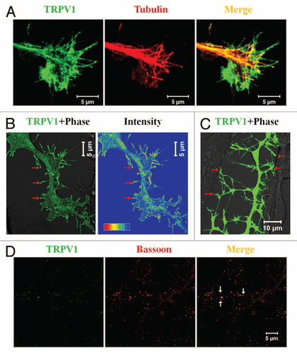
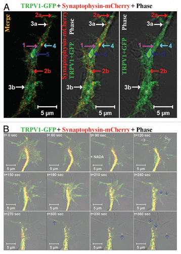
Figure 2 Distribution and movement of TRPV1 via different types of synaptic vesicles. (A) Distribution of TRPV1-GFP in different population within a neurite. Show are the live cell confocal images of a neurite developed from F11 cell expressing TRPV1-GFP (green) and Synaptophysin-mCherry (red). TRPV1-GFP is detected in different groups: (1) A diffused pattern which is mostly cytoplasmic and recovers fast from all sides after photo-bleaching (indicated by pink arrow), (2a and b) medium- and small-sized particles that move uninterruptedly with an average speed and travels long distance (indicated by red arrows respectively), (3a and b) the small particles located at the base of the existing filopodia and can move fast within the filopodial structures after activation (indicated by white arrows respectively), (4) medium-sized (indicated by blue arrow) particle which mostly stay at the inner side of the neuritic membrane but do not move. This population uptake FM4-64 dye rapidly, (5) some of the much bigger entities (indicated by a dark blue arrow and a marked region) generally remain immobile for a long time and show only flickering. These bigger particles can move all of a sudden to a short distance and very fast. Some of these entities can be categorized as cytoplasmic transport packets (CTP) as these entities fit well with the properties of CTPs. Scale bar 5 µm. (B) Shown are the live cell confocal images of a growth cone developed from F11 cell expressing TRPV1-GFP (green) and Synaptophysin-mCherry (red). Note that activation of F11 cells by NADA (at 90 sec time frame), an endogenous stimulus for TRPV1 results in rapid translocation of TRPV1-GFP to the plasma membrane (indicated by white arrows at 120 sec) resulting in sudden increase in the intensity of TRPV1-GFP at the of growth cone membrane. Activation also results in selective co-migration of TRPV1-GFP-containing pre-synaptic vesicles to filopodial structures (indicated by blue arrows). Often the movement of these vesicles follows a distinct route. So far the exact signaling events and the molecular mechanisms which regulate this translocation to membrane and movement of vesicles are not known. Scale bar 5 µm.
Addendum to: