Abstract
Background: Neurothekeomas are rare, benign neoplasms, typically occurring in younger patients with a remarkable predilection for the female population. The nomenclature and derivation of these tumors is controversial. The rarity of this unusual skin tumor in daily routine histopathologic findings prompted the following report.
Observation: We report a 17-year-old girl with an asymptomatic nodule at the inner angle of the left eye with slow progression in size within 12 months to 1 cm in diameter. Multiple treatments with laser surgery were performed and yielded no persistent remission of the tumor. Histopathologic examination revealed a non-encapsulated dermal tumor, composed of multiple, closely situated medium-sized nodules, separated by a myxoid collagen-rich stroma, without epidermal alteration. In summary, due to the histoarchitecture and immunoprofile of the tumor, a mixed-type cellular neurothekeoma was diagnosed.
Conclusion:
We think that it is important to be aware of these uncommon soft tissue lesions and the pitfalls of mixed-type neurothekeomas that often cause diagnostic problems. The aim of this report is to help to avoid misdiagnoses of malignant mesenchymal tumors with serious consequences, including extensive surgical therapy or radiation.
Neurothekeomas are rare, benign neoplasms, typically occurring in younger patients with a remarkable predilection for the female population. The nomenclature and derivation of these tumors is controversial. Specifically, the origin of neurothekeomas has been ascribed to peripheral nerve sheath cells, epitheloid variants of dermatofibromas, and even pilar leiomyomas.Citation1–Citation3 The rarity of this unusual skin tumor in daily routine histopathologic findings prompted the following report.Citation2
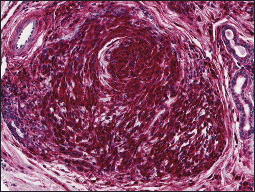
A 17-year-old girl was referred to our institution with an asymptomatic, unilateral, papillomatous, pale, erythematous nodule at the inner angle of the left eye (). She described a slow progression of the tumor size within 12 months to 1 cm in diameter at the first dermatologic consultation. There was no history of trauma or insect bites. Prior to the primary consultation in our department, multiple treatments with laser surgery were performed and yielded no persistent remission of the tumor. Histopathologic examination revealed a non-encapsulated dermal tumor, composed of multiple, closely situated medium-sized nodules, separated by a myxoid collagen-rich stroma (). No epidermal alteration or infiltration of the subcutaneous tissue was observed. Immunohistochemical studies displayed strong immunoreactivity for NKI/C3 () and smooth-muscle actin, while neuronal markers, such as neuron-specific enolase and S100 were non-reactive.Citation7 Melan-A, HMB-45, CD34, pan-cytokeratin and CD68 were not detectable in the tumor cells. In summary, due to the histoarchitecture and immunoprofile of the tumor, a mixed-type cellular neurothekeoma was diagnosed, with the classical myxoid fraction remarkably lacking S100 reactivity.
Historically, three subsets of neurothekeomas have been distinguished: myxoid, cellular and mixed.Citation4,Citation5 The classic, or myxoid type was first described by Harkin and Reed in 1969 as a “myxoma of the nerve sheath,” occurring primarily in adults with a predilection for the head and neck region of females. Clinically, neurothekeomas are solitary tender nodules, often mistaken as dermal nevi, cysts or adnexal neoplasms. The histologic findings are lobular-to-plexiform dermal tumor masses composed of spindle-to-stellate-shaped cells, with a striking myxoid stroma. Immunoreactivity is strong for S100. Cellular neurothekeomas first described in 1986 by Rosati andCitation5 are commonly seen in younger people in the head and neck region. Histopathologically, these dermal tumors are poorlycircumscribed, typically being composed of fascicles of spindle-shaped-to-epitheloid tumor cells. Intriguingly, these cells are not immunoreactive for S100, but have a strong reactivity for NKIC3 and even strong reactivity for smooth muscle actin, as in the case presented herein.Citation2,Citation5
The so-called “mixed-type” of neurothekeomas shows overlapping features of both variants.Citation2,Citation6 The immunohistochemistry in these cases often show confusing features with an irregular or lacking reactivity to S100 and smooth muscle actin,Citation4,Citation5 as shown herein. The discussion continues from which cell line neurothekeomas are derived. Until now, it has been widely accepted that there is no real relationship to nerve sheath myxomas, as introduced by Gallager and Helwig in 1980.Citation4 Calonje, et al.Citation1 have argued that cellular neurothekeomas are an epitheloid variant of pilar leiomyomas due to their smooth muscle actinpositive immunophenotype.Citation1 Zelger, et al.Citation3 consider these tumors as epitheloid variants of dermatofibromas. The differential diagnoses include myxoid malignant fibrous histiocytomas and myxoid neurofibromas. Melanocytic lesions have to be excluded by well-known sets of classical immunomarkers. Although neurothekeomas are benign neoplasms with a little tendency for local recurrence, total excision of these tumors is warranted. As shown in our case, treatment with laser surgery and multiple local excisions is not able to cure the neoplasm ultimately; for this reason, we performed a radical micrographic controlled excision of the entire tumor.
We think that it is important to be aware of these uncommon soft tissue lesions and the pitfalls of mixed-type neurothekeomas that often cause diagnostic problems. The aim of this report is to help to avoid misdiagnoses of malignant mesenchymal tumors with serious consequences, including extensive surgical therapy or radiation.
Figures and Tables
Figure 1 A 17-year-old girl with an asymptomatic, unilateral, papillomatous, pale, erythematous nodule at the inner angle of the left eye
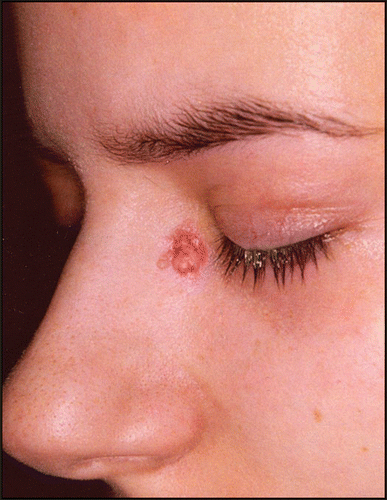
Figure 2 HE sections displaying solid nodules of spindle-shaped cells in a myxoid stroma.
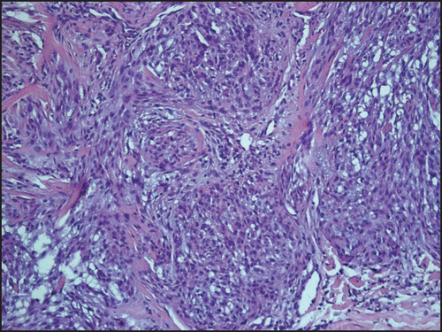
Figure 3 Spindle-shape cell formations express NKI/C3.
Acknowledgements
We thank Anne Kerber for excellent histological and immunohistological stainings.
References
- Calonje E, Wilson-Jones E, Smith NP, Fletcher CD. Cellular ‘neurothekeoma’: an epithelioid variant of pilar leiomyoma? Morphological and immunohistochemical analysis of a series. Histopathology 1992; 20:397 - 404
- Mentzel T. Cutaneous neural neoplasms—an update. Pathologe 1999; 20:98 - 109
- Zelger BG, Zelger B. Cellular ‘neurothekeoma’: an epithelioid variant of dermatofibroma?. Histopathology 1998; 32:414 - 422
- Fetsch JF, Laskin WB, Hallmann JR, Lupton GP, et al. Neurothekeoma: an analysis of 178 tumors with detailed immunohistochemical data and long-term patient follow-up information. Am J Surg Pathol 2007; 31:1103 - 1114
- Hornick JL, Fletcher CD. Cellular neurothekeoma: detailed characterization in a series of 133 cases. Am J Surg Pathol 2007; 31:329 - 340
- Yang YW, Shih IH, Huang YH, Kuo TT, et al. Mixed-type neurothekeoma presenting with an unusual clinical appearance of multiple satellite lesions on the back. Dermatol Surg 2005; 31:720 - 722
- Page RN, King R, Mihm MCJR, et al. Microphthalmia transcription factor and NKI/C3 expression in cellular neurothekeoma. Mod Pathol 2004; 17:230 - 234