
Abstract
Eccrine angiomatous hamartoma (EAH) is a rare, benign cutaneous lesion characterized histologically by a proliferation of eccrine glands and vascular structures—generally capillaries—in the middle and deep dermis. Sudden enlargement of EAH lesions with or without pain has been noted during puberty and pregnancy and has been attributed to hormonal stimulation. We herein describe a case of EAH that became symptomatic in an adolescent girl. A 13-year-old girl presented with pain associated with a sudden enlargement of a previously asymptomatic swelling on her right second toe. She had an 8-year history of an asymptomatic swelling on her right second toe, and the symptoms appeared approximately 1 year after menarche. Physical examination revealed swelling of the plantar surface of her right second toe. The overlying surface was erythematous with a small amount of fine scales. The biopsied tissue showed a nodular proliferation of eccrine glands intimately admixed with numerous small vessels in the deep dermis and subcutaneous fat tissue. Mucin deposition was present in the stroma surrounding the proliferating eccrine coils and ducts and in the upper dermis. A diagnosis of EAH was made. We suggest that hormonal changes during puberty may have played a role in the rapid growth and pain in the present case.
Figures and Tables
Figure 1 Clinical appearance of the skin lesion. (A–C) Swelling of the plantar surface was apparent on the right second toe. The overlying surface was erythematous with a small amount of fine scales. Histologically, the epidermis showed acanthosis with hypertrophy of the granular layer and mild elongation of rete ridges. (D) A mild, perivascular inflammatory infiltration in the upper dermis was seen (hematoxylin and eosin, original magnification x40). (E) A nodular proliferation of normally structured eccrine coils and ducts in the deep dermis and subcutaneous fat tissue. Some of the ducts show mild dilation. Extravasation of erythrocytes was also noted around the proliferation of eccrine glands (hematoxylin and eosin, original magnification x200). (F) Proliferation of small capillary-sized vessels in the stroma surrounding the eccrine glands in the subcutaneous fat tissues (hematoxylin and eosin, original magnification x200).
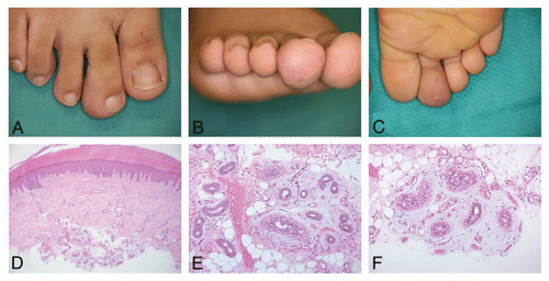
Table 1 Painful skin tumors/malformations