
Abstract
The ability of environmental factors to shape health and disease involves epigenetic mechanisms that mediate gene-environment interactions. Metastable epiallele genes are variably expressed in genetically identical individuals due to epigenetic modifications established during early development. DNA methylation within metastable epialleles is stochastic due to probabilistic reprogramming of epigenetic marks during embryogenesis. Maternal nutrition and environment have been shown to affect metastable epiallele methylation patterns and subsequent adult phenotype. Little is known, however, about the role of histone modifications in influencing metastable epiallele expression and phenotypic variation. Utilizing chromatin immunoprecipitation followed by qPCR, we observe variable histone patterns in the 5’ long terminal repeat (LTR) of the murine viable yellow agouti (Avy) metastable epiallele. This region contains 6 CpG sites, which are variably methylated in isogenic Avy/a offspring. Yellow mice, which are hypomethylated at the Avy LTR and exhibit constitutive ectopic expression of agouti (a), also display enrichment of H3 and H4 di-acetylation (p=0.08 and 0.09, respectively). Pseudoagouti mice, in which Avy hypermethylation is thought to silence ectopic expression, exhibit enrichment of H4K20 tri-methylation (p=0.01). No differences are observed for H3K4 tri-methylation (p=0.7), a modification often enriched in the promoter of active genes. These results show for the first time the presence of variable histone modifications at a metastable epiallele, indicating that DNA methylation acts in concert with histone modifications to affect inter-individual variation of metastable epiallele expression. Therefore, the potential for environmental factors to influence histone modifications, in addition to DNA methylation, should be addressed in environmental epigenomic studies.
Acknowledgements
This work was supported by NIH grants ES017524, ES13053, ES08823 and ES015165. The authors would like to thank Mary Ellen Koran and Dale Huang for technical assistance.
Figures and Tables
Figure 1 The Avy metastable epiallele. (A) The viable yellow agouti (Avy) allele contains a contra-oriented IAP insertion within pseudoexon 1A (PS1A) of the Agouti gene. A cryptic promoter (short arrow labeled Avy) drives constitutive ectopic Agouti expression. Transcription of the Agouti gene normally initiates from a developmentally regulated hair-cycle specific promoter in exon 2 (short arrow labeled A, a). (B) Genetically identical Avy/a offspring representing the yellow and pseudoagouti coat color phenotypes are shown. * indicates 5′LTR of the Avy IAP region and **represents non-IAP genomic PS1A region.
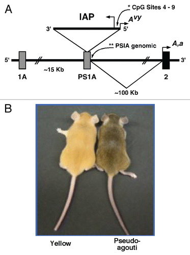
Figure 2 Chromatin precipitation for acetylated histones H3 and H4 in the 5′ LTR of the IAP and in PS1A. Binding activity was calculated as percent of pre-immunoprecipitated input DNA as represented by 2−ΔC(t) X100. (A) DNA precipitated by H3 di-acetylation antibody is enriched in yellow versus pseudoagouti Avy/a mice (p = 0.09; n = 6 per group) within the 5′ LTR of the IAP. (B) DNA precipitated by H4 di-acetylation antibody is enriched in yellow versus pseudoagouti Avy/a mice (p = 0.08; n = 3 per group) within the 5′ LTR of the IAP. (C) DNA precipitated by H3 di-acetylation antibody does not vary by coat color within the PS1A genomic region (p = 0.4; n = 6 per group). (D) DNA precipitated by H4 di-acetylation antibody does not differ by coat color within the genomic PS1A region (p = 0.4; n = 3 per group). * indicates significance at the 0.09 level.
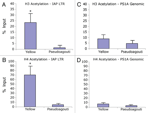
Figure 3 Chromatin precipitation for methylated histones in the 5′ LTR of the IAP and in PS1A. Binding activity was calculated as percent of pre-immunoprecipitated input DNA as represented by 2−ΔC(t) X100. (A) DNA precipitated by H3K4 tri-methylation antibody is not enriched in yellow versus pseudoagouti Avy/a mice (p = 0.7; n = 6 per group) within the 5′ LTR of the IAP. (B) DNA precipitated by H4K20 tri-methylation antibody is enriched in pseudoagouti compared to yellow Avy/a mice (p = 0.01; n = 6 per group) within the 5′ LTR of the IAP. (C) DNA precipitated by H3K4 tri-methylation antibody does not vary by coat color within the PS1A genomic region (p = 0.9; n = 5 per group). (D) DNA precipitated by H4K20 tri-methylation antibody does not differ by coat color within the genomic PS1A region (p = 0.8; n = 5 per group). ** indicates significance at the 0.01 level.
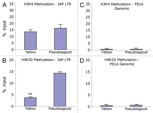
Table 1 DNA methylation and Agouti (a) expression levels by histone and coat color status
Table 2 PCR and pyrosequencing primers