Abstract
MicroRNAs (miRNAs) are short non-coding RNA molecules that regulate post-transcriptional gene expression. They influence a wide range of physiological functions, including neuronal processes, and are regulated by various mechanisms, such as DNA methylation. This epigenetic mark is recognized by transcriptional regulators such as the methyl CpG binding protein Mecp2. Rett syndrome is a complex neurological disorder that has been associated with mutations in the gene coding for Mecp2. Thus, we examined the possible miRNA misregulation caused by Mecp2 absence in a mouse model of Rett syndrome. Using miRNA expression microarrays, we observed that the brain of Rett syndrome mice undergoes a disruption of the expression profiles of miRNAs. Among the significantly altered miRNAs (26%, 65 of 245), overall downregulation of these transcripts was the most common feature (71%), whilst the remaining 30% were upregulated. Further validation by quantitative RT-PCR demonstrated that the most commonly disrupted miRNAs were miR-146a, miR-146b, miR-130, miR-122a, miR-342 and miR-409 (downregulated), and miR-29b, miR329, miR-199b, miR-382, miR-296, miR-221 and miR-92 (upregulated). Most importantly, transfection of miR-146a in a neuroblastoma cell line caused the downregulation of IL-1 receptor-associated kinase 1 (Irak1) levels, suggesting that the identified defect of miR-146a in Rett syndrome mice brains might be responsible for the observed upregulation of Irak1 in this model of the human disease. Overall, we provide another level of molecular deregulation occurring in Rett syndrome that might be useful for understanding the disease and for designing targeted therapies.
Acknowledgements
We are grateful to the Catalan and Valencian Rett Syndrome Associations for their support. R.G.U. is funded by the Comunidad de Madrid FPI Programme. The research leading to these results has received funding from the European Community's Sixth Framework Programme (FP7/2007-2013) under grant agreement n° PITN-GA-2009-238242—DISCHROM project, from the Instituto de Salud Carlos III—Ministerio de Sanidad y Consumo Proyecto FIS (Fondo Investigación Sanitaria, Spain) through the E-RARE EuroRETT network, from the Fondation Lejeune (France) and from Grant SAF2007-6-8.
Figures and Tables
Figure 1 miRNAs upregulated and downregulated in the brain of a mouse model for Rett syndrome. Validation by quantitative RT-PCR provided the most commonly disrupted miRNAs. Example of the expression of the miR-29b in pairs wild-type/Mecp2 null brain showing upregulation of the transcript in the disease-associated tissue.
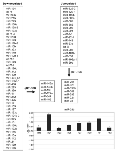
Figure 2 DNA methylation and Mecp2 occupancy analysis for the 5′-end CpG islands of miR-29b, miR-199b, miR-146a and miR-146b. (A) Bisulfite genomic sequencing of multiple clones. Black squares, methylated cytosines; white squares, unmethylated cystosines. Vertical black arrow, transcription start sites of the miRNA. Black arrows, bisulfite genomic sequencing primers; white arrows, chromatin immunoprecipitation primers. (B) Quantitative chromatin immunoprecipitation (ChIP) assays. Mecp2 occupancy is shown in the wild-type brain for the miR-29b-1, miR-146a and miR-146b CpG islands. IgG and H3 are used as negative and positive controls for the ChIP assay.
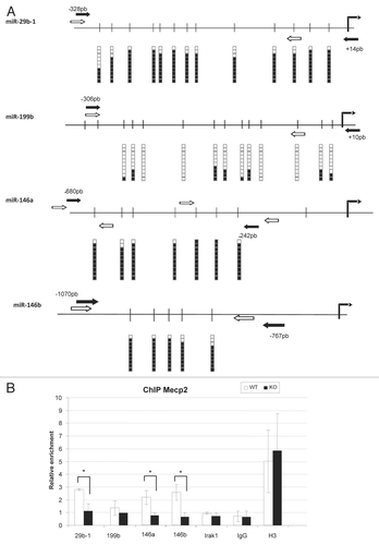
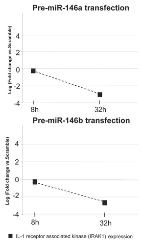
Figure 3 Downstream targets upon miRNA deregulation in Rett syndrome. Transfection of the precursor molecules of miR-146a and miR-146b downregulates IL-1 receptor-associated kinase 1 (Irak1) expression in the mice neuroblastoma cell line Neuro-2a.