Abstract
5-Hydroxymethyl-cytosine (5-hmC) is a form of modified cytosine, which has recently attracted a considerable attention due to its potential role in transcriptional regulation. According to several reports 5-hydroxymethyl-cytosine distribution is tissue-specific in mammals. Thus, 5-hmC is enriched in embryonic cell populations and in adult neuronal tissue. Here, we describe a novel method of semi-quantitative immunohistochemical detection of 5-hmC and utilize it to assess the levels of this modification in amphibian tissues. We show that, similar to mammalian embryos, 5-hmC is enriched in axolotl tadpoles compared with adult tissues. Our data demonstrate that 5-hmC distribution is tissue-specific in amphibians, and that strong 5-hmC enrichment in neuronal cells is conserved between amphibians and mammals. In addition, we identify 5-hmC-enriched cell populations that are distributed in amphibian skin and connective tissue in a mosaic manner. Our results illustrate that immunochemistry can be successfully used not only for spatial identification of cells enriched with 5-hmC, but also for the semi-quantitative assessment of the levels of this epigenetic modification in single cells of different tissues.
Disclosure of Potential Conflicts of Interest
No potential conflicts of interest were disclosed.
Acknowledgments
We thank Lorraine Young (STEM, University of Nottingham) for supporting this study and Alexander Kondrashev (University of Nottingham) for help. The authors declare no conflict of interest.
Figures and Tables
Figure 1 (A) A bootstrap consensus tree inferred using Neighbor-Joining from 100 replicates showing the relationships between Xenopus, zebrafish, chick, mouse and human Tet protein sequences. Although the tree gives the appearance of rooting, it is essentially unrooted. The root is placed at the midpoint of the tree to simplify the presentation. The percentages of replicate trees in which the associated taxa clustered together are shown next to the branches. (B) The developmental stage and tissue distribution of Xenopus tropicalis Tet2 and 3 transcripts is determined from NCBI UniGene EST profiles (Tet2:Str.52041 Tet3:Str.53063). Transcript counts are reported in TPM (transcripts per million). (C–G) Immunohistochemical detection of 5-hmC and 5-mC in the tissues of axolotl tadpole. Immunostaining for 5-hmC, 5-mC and merge views are shown. The locations of (D–G) views are indicated with dotted squares on (C).
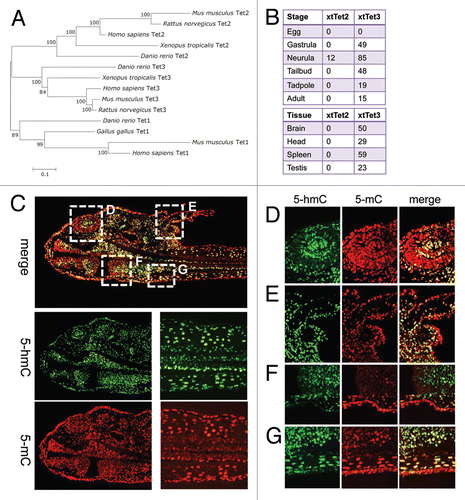
Figure 2 The distribution of 5-hydroxymethyl-cytosine in adult Xenopus (A) and axolotl (B and C) tissues. 5-hydroxymethylcytsoine and 5-methyl-cytosine have been detected in indicated tissues using 1:5,000 dilution of anti-5-hmC antibody. Immunostaining for 5-hmC, 5-mC and merge views are shown. Skel muscle-skeletal muscle. Scale bars are 20 µm.
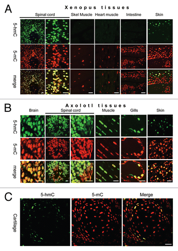
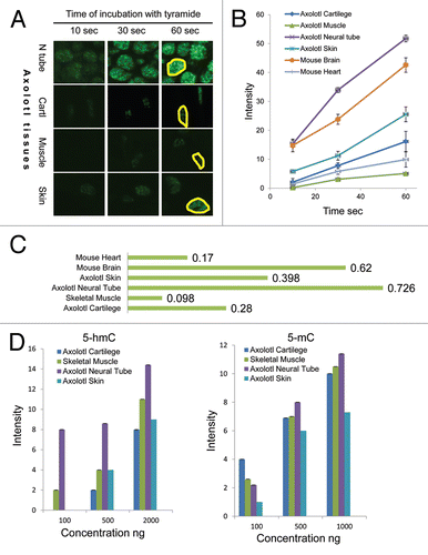
Figure 3 The semi-quantitative assessment of 5-hmC distribution in adult axolotl tissues. (A) 5-hmC immunostaining signal using 1:50,000 dilution of primary antibody at indicated times of incubation with tyramide on sections of axolotl spinal cord (neural tube, N tube), skeletal muscle (Muscle), skin and connective tissue adjacent to cartilage (Cartl). Adjacent sections were stained in parallel at identical conditions with different times of incubation with tyramide. The exposures are identical for all the presented pictures. Examples of regions, which were used for signal quantification, are shown with yellow-line shapes. (B) The progress curves of peroxidase reactions produced by quantification of immunostaining data for different axolotl and mouse tissues. “Skin” and “cartilage” refers to 5-hmC enriched cells found in these tissues. Most of skin and cartilage cells do not exhibit any detectable staining at any times of incubation with tyramide (A). (C) The velocities of peroxidase reactions for different axolotl and mouse tissues (indicated). (D) The quantification of the dot-blot results performed with a total DNA derived from the indicated axolotl tissues using anti-5-hmC (5-hmC) and anti-5-mC (5-mC) antibodies. The graphs show the dependence of the dot-blot signal intensity of different concentrations of total DNA (indicated).