
Abstract
Expression of recombinant vaccine antigens and monoclonal antibodies using plant viral vectors has developed extensively during the past several years. The approach benefits from high yields of recombinant protein obtained within days after transient delivery of viral vectors to leaves of Nicotiana benthamiana, a tobacco relative. Modified viral genomes of both RNA and DNA viruses have been created. Geminiviruses such as bean yellow dwarf virus (BeYDV) have a small, single stranded DNA genome that replicates in the nucleus of an infected plant cell, using the cellular DNA synthesis apparatus and a virus-encoded replication initiator protein (Rep). BeYDV-derived expression vectors contain deletions of the viral genes encoding coat and movement proteins and insertion of an expression cassette for a protein of interest. Delivery of the geminiviral vector to leaf cells via Agrobacterium-mediated delivery produces very high levels of recombinant DNA that can act as a transcription template, yielding high levels of mRNA for the protein of interest. Several vaccine antigens, including Norwalk virus capsid protein and hepatitis B core antigen, were expressed using the BeYDV vector at levels up to 1mg per g of leaf mass. BeYDV replicons can be stacked in the same vector molecule by linking them in tandem, which enables production of multi-subunit proteins like monoclonal antibody (mAb) heavy and light chains. The protective mAb 6D8 against Ebola virus was produced at 0.5mg per g of leaf mass. Multi-replicon vectors could be conveniently used to produce protein complexes, e.g. virus-like particles that require two or more subunits.
Acknowledgements
This work was supported by NIH grants U19 AI066332 to H. Mason and U01 AI075549 to Q. Chen.
Figures and Tables
Figure 1 Bean yellow dwarf virus genome structure. The genome is circular single-stranded DNA of 2,561 nt in length. The long intergenic region (LIR) contains a bidirectional promoter that drives transcription of the virion sense genes (V1, movement protein; V2, coat protein) and the complimentary sense genes (C1, RepA; C1/C2 with spliced intron, Rep protein). The LIR also contains a stem-loop structure that is the site of nicking by Rep protein. The short intergenic region contains polyadenylation signals for the protein-coding genes.
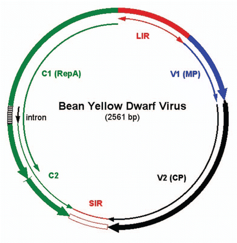
Figure 2 Structure of BeYDV replicon encoding GFP. The T-DNA construct (top) is flanked by left border (LB) and right border (RB) sequences that delineate the DNA to be transferred by Agrobacterium to the plant cells. The replicon is flanked by two LIR elements (red blocks), and the GFP expression cassette is inserted in place of the V1 and V2 genes (), between the one LIR and the SIR (yellow oval). The Rep/RepA (C1/C2) gene is located between the SIR and the other LIR, in its normal position in the BeYDV genome (). Once delivered into plant cells, T-DNA is targeted to the nucleus, where transcription of C1/C2 produces Rep mRNA, which is transported to the cytoplasm and translated to make Rep protein. The Rep protein is targeted back to the nucleus, where it nicks the LIR stem-loops in the T-DNA to release a single-stranded DNA segment. The DNA is then copied to make double-stranded DNA, and rolling circle replication occurs to produce very high copy number of replicons. In double stranded replicative form, the DNA is a template for mRNA transcription and yields GFP mRNA in abundance. 35S/TEV, CaMV 35S promoter linked to the tobacco etch virus 5′UTR; GFP, coding sequence for green fluorescent protein; VSP 3′, soybean vspB 3′ flanking region with transcription termination signals.
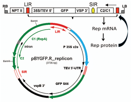
Figure 3 BeYDV replicon vectors available for use. pBYR1 and pBYR2 are T-DNA vectors for use with Agrobacterium DNA delivery, whose structure is similar to that of pBYGFP.RCitation19 (). The GFP coding sequence is replaced by a polylinker with several unique restriction sites (bold font). Both expression cassettes use the CaMV 35S promoter with duplicated enhancer. pBYR1 has the TEV 5′ UTR and vspB 3′ region, while pBYR2 has the TMV 5′ UTR and the extensin (Ext) 3′ region. Both pBYR1 and pBYR2 have been modified to replace the NPTII expression cassette with one encoding the silencing suppressor p19 from TBSV, to produce pBYR1p19 and pBYR2p19.
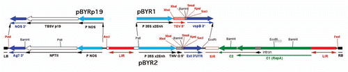
Figure 4 West Nile virus chimeric VLP produced with BeYDV replicon in N. benthamiana plants. (A) Expression of HBc-DIII. Leaves were infiltrated with Agrobacterium transformed with the HBc-DIII construct. Proteins were extracted on days 3 to 7 post Agro-infiltration (DPI) and were analyzed with an ELISA that detects HBcAg.Citation19 Data are mean ± standard error (SE M) of samples from three independent infiltration experiments. (B) Electron microscopy of purified HBc-DIII. HBc-DIII was purified and subject to negative staining with 0.2% aqueous uranyl acetate, and transmission electron microscopy with a Philips CM-12 microscope. (C and D) Western blot analysis of HBc-DIII. Leaf proteins were separated on 12% SDS-PAGE gels under reducing condition and blotted onto PVDF membranes. The membranes were incubated with an anti-HBc antibodyCitation19 (C) or an anti-DIII antibodyCitation39 (D). Lane 1, Protein sample extracted from leaves infiltrated with the HBc-DIII construct; lane 2, Extract from un-infiltrated leaves.
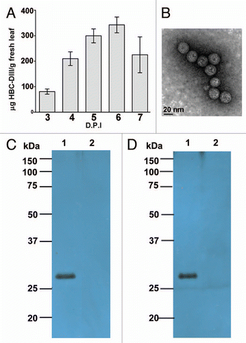
Figure 5 Dual BeYDV replicon vector for co-expression of two genes. The T-DNA construct (top) contains two replicons linked in tandem, with each flanked by an LIR (red blocks). The vector shown is pBY-HL(6D8).R, which drives expression of the heavy and light chains of the anti-Ebola virus mAb 6D8.Citation9 The central LIR functions for both replicons to allow release of two separate single stranded DNA molecules that can enter the rolling circle replication process. The SIR elements are indicated by the yellow ovals. Both replicons are amplified non-competitively to high copy numbers in the nucleus, and generate abundant mRNA's for the heavy and light chains. The pBYK3R replicon (right) contains the C1/C2 gene in its normal position in the viral genome (), and produces Rep mRNA to make Rep protein, which can then act upon both replicons to facilitate their replication. The pBYH2kdel replicon has a large segment of C1/C2 deleted (BamHI fragment, ), since C1/C2 transcription from only one replicon is required. LB and RB, left and right borders of the T-DNA; NPTII, expression cassette for NPTII (facilitates selection of stable transgenic plant lines, but not required for transient expression); 35S/TEV 5′, CaMV 35S promoter linked to tobacco etch virus 5′UTR; H(6D8), coding sequence for mAb 6D8 heavy chain (“H2-SEKDEL” in the replicon below); VSP 3′, soybean vspB 3′ flanking region; 35S/TMV 5′, CaMV 35S promoter linked to TMV 5′UTR; L(6D8), coding sequence for mAb 6D8 light chain (“K chain” in the replicon below); rbcS 3′, tobacco rbcS gene 3′ flanking region; C2/C1, Rep/RepA coding sequence.
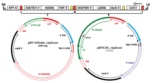
Table 1 Proteins expressed using geminiviral replicons