
Abstract
Tumor necrosis factor (TNF) signals through two membrane receptors, TNFR1 and TNFR2, and TNFR1 is known to be the major pathogenic mediator of chronic and acute inflammatory diseases. Present clinical intervention is based on neutralization of the ligand TNF. Selective inhibition of TNF receptor 1 (TNFR1) provides an alternative opportunity to neutralize the pro-inflammatory activity of TNF while maintaining the advantageous immunological responses mediated by TNFR2, including immune regulation, tissue homeostasis and neuroprotection. We recently humanized a mouse anti-human TNFR1 monoclonal antibody exhibiting TNFR1-neutralizing activity. This humanized antibody has been converted into an IgG1 molecule (ATROSAB) containing a modified Fc region previously demonstrated to have greatly reduced effector functions. Purified ATROSAB, produced in CHO cells, showed strong binding to human and rhesus TNFR1-Fc fusion protein and mouse embryonic fibroblasts transfected with a recombinant TNFR1 fusion protein with an affinity identical to the parental mouse antibody H398. Using chimeric human/mouse TNFR1 molecules, the epitope of ATROSAB was mapped to the N-terminal region (amino acid residues 1-70) comprising the first cysteine-rich domain (CRD1) and the A1 sub-domain of CRD2. In vitro, ATROSAB inhibited typical TNF-mediated responses like apoptosis induction and activation of NFκB-dependent gene expression such as IL-6 and IL-8 production. These findings open the way to further analyze the therapeutic activity of ATROSAB in relevant disease models in non-human primates.
Acknowledgements
This work was supported by a grant from EC FP6, Project NeuroproMiSe, contract # LSHM-CT-2005-018637.
Figures and Tables
Figure 1 Characterization of ATROSAB. (A) SDS-PAGE analysis of purified ATROSAB (4 µg/lane, Coomassie staining) analyzed under non-reducing (1) or reducing (2) conditions. (B) Size exclusion chromato-graphy of ATROSAB (the position of standard proteins is indicated). (C) ELISA of ATROSAB and H398 for binding to human TNFR1-Fc.
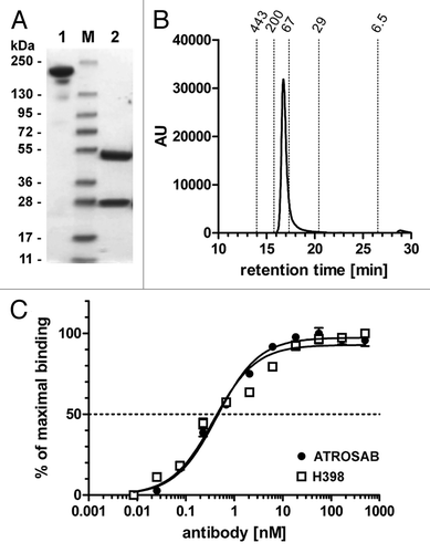
Figure 2 Flow cytometry analysis of binding of ATROSAB to mouse embryonic fibroblasts (MEF) transfected with human TNFR1-Fas (A) or human TNFR2-Fas (B). (C) Titration of binding of ATROSAB and H398 to MEF-TNFR1-Fas (n = 3).
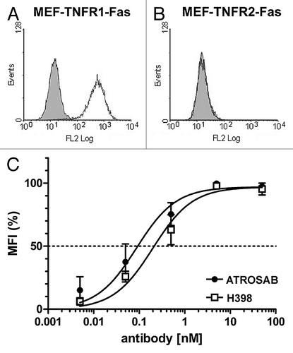
Figure 3 (A) SDS-PAGE analysis of purified human TNFR1-Fc (1 and 4), mouse TNFR1-Fc (2 and 5) and rhesus TNFR1-Fc (3 and 6) (4 µg/lane, Coomassie staining) analyzed under reducing (1–3) and non-reducing (4–6) conditions. (B) ELISA of binding of ATROSAB and H398 (5 µg/ml) to purified human TNFR1-Fc, rhesus TNFR1-Fc and mouse TNFR1-Fc (100 ng/well). Binding was detected by HRP-conjugated anti-moIgG (Fc-specific) antibody or anti-human Fab antibody, respectively. Binding of an anti-human Fc antibody (anti-Fc) was included as coating control.
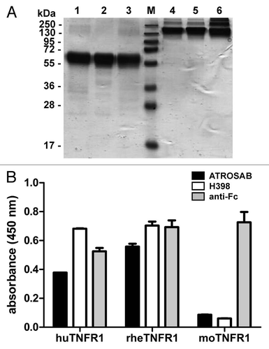
Figure 4 Determination of affinity of H398 and ATROSAB for binding to human and rhesus TNFR1-Fc by quartz crystal microbalance (QCM) measurements. (A) Binding of H398 to human TNFR1-Fc, (B) binding of ATROSAB to human TNFR1-Fc, (C) binding of H398 to rhesus TNFR1-Fc and (D) binding of ATROSAB to rhesus TNFR1-Fc.
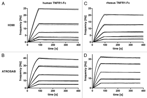
Figure 5 Inhibition of TNF-mediated cytotoxicity (1.25 ng/ml TNF) on Kym-1 cells by ATROSAB and H398. Cells were analyzed after 6 h by crystal violet staining (n = 3). Maximum (10% viability of control) and half maximum (55% viability of control) are displayed in dotted lines.
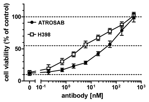
Figure 6 Inhibition of IL-6 and IL-8 secretion induced by TNF by ATROSAB and H398. HeLa cells (A) or HT1080 cells (B) were incubated with TNF (1 ng/ml) and increasing concentrations of ATROSAB or H398 and cytokine secretion were determined by ELISA (n = 3). Human IgG (huIgG) was incluced as negative control. In the same way, effects of antibodies on cytokine secretion in the absence of TNF were determined. Compared with TNF, both antibodies had only marginal effects on IL-6 (C) and IL-8 (D) secretion.
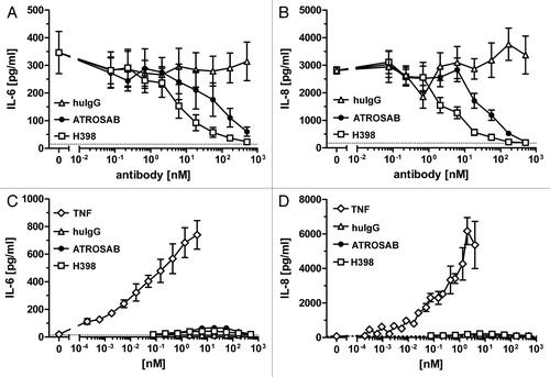
Figure 7 Plasma half-lives of ATROSAB and H398 after a single dose i.v. injection (25 µg) into CD1 mice. Serum concentrations of antibodies were determined by ELISA.
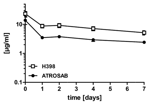
Figure 8 (A) Epitope mapping of ATROSAB and H398 using wild-type and chimeric human/mouse TNFR1-Fc fusion proteins. Antibodies (0.1 nM) were analyzed by ELISA for binding to the TNFR1-Fc fusion proteins. His-tagged human TNF (huTNF) was included as control. (B) Sequence comparison of the identified epitope region (aa 1–70) of human (huTNFR1), mouse (moTNFR1) and rhesus (rhTNFR1) TNFR1. Cysteine residues are marked with grey boxes and the two positions (P23, Q24) analyzed by site-directed mutagenesis are marked by asterisks.
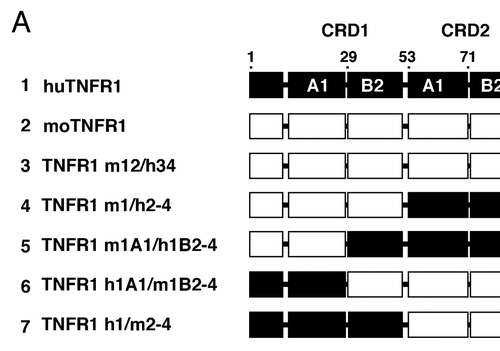
Figure 9 (A) Structure of TNF (red) bound to TNFR1 (blue). The identified epitope region is marked in green. (B) A single TNFR1 chain. The two positions (P23, Q24) identified by mutagenesis to contribute to binding of ATROSAB and H398 are highlighted in dark green.
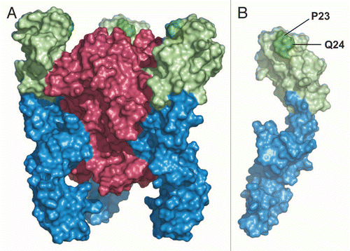
Table 1 Binding kinetics of H398 and ATROSAB