Abstract
The neonatal Fc receptor (FcRn) regulates IgG and albumin homeostasis, mediates maternal IgG transport, is active in phagocytosis and delivers antigen for presentation. We have previously shown that transgenic (tg) mice that have been created to overexpress bovine FcRn (bFcRn) demonstrate increased half-life of mouse IgG, significantly increased antigen-specific IgG in serum and augmented expansion of antigen-specific B cells and plasma cells after immunization. One of the interesting questions surrounding this enhanced immune response is whether these tg mice could effectively induce immune response to weakly immunogenic antigens. To address this question, we immunized these bFcRn tg mice with a conserved hemagglutinin subunit 2 (HA2)-based synthetic peptide that was recently found to be effectively targeted by neutralizing antibodies. Using an ELISA system, we found that, whereas wild-type mice showed a weak immune response and developed only a de minimis amount of antibody against the epitope, FcRn over-expressing animals mounted a robust reaction expressed in specific antibody titers on day 28 that continued to rise through day 50. Consistent with our previous data, the enhanced immune response resulting from the FcRn overexpression was also associated with a substantial increase in the number of spleen derived B cells, dendritic cells, granulocytes and plasma cells. Based on this evidence, we propose that tg mice that overexpress bFcRn offer major advantages in monoclonal antibody production because the tg mice would allow the generation of antibodies (hybridomas) to weakly immunogenic antigens that otherwise would be difficult or even impossible to make.
Acknowledgements
Supported by the grant OM-00117/2008 from the Hungarian National Office for Research and Technology and ImmunoGenes, Hungary.
Conflict of Interest
I.K. is one of the founders, A.V. and J.C. are researchers at ImmunoGenes Kft, a spin-off company from University Eotvos Lorand, Budapest and Agricultural Biotechnology Center, Godollo, specialized in the generation of transgenic animals for the production of polyclonal and monoclonal antibodies.
Figures and Tables
Figure 1 Antigen selection for immunizing bFcRn tg mice. (A) Crystal structure of Fab CR6261 in complex with the 1918 H1N1 influenza virus hemagglutinin.Citation10 The HA1 globular head is located at the top of the figure. One HA/Fab protomer is colored with HA1 in purple, HA2 in blue, Fab heavy chain in brown and Fab light chain in green. Yellow balls on HA2 indicate residues that are involved, while grey balls indicate those residues that are not involved in binding to CR6261 of the selected amino acids (41–57), respectively. Figure was created by using Cn3D of NCBICitation32,Citation33 using the crystal structure 3GBN (A).Citation10 This segment is composed of highly conserved amino acids 41–57 from the HA2 of different hemagglutin subtypes; white letters on a black background indicate a residue conserved in all five HAs; black letters on a gray background indicate a residue conserved in four of five HAs or less stringent conservation. Asterisk (*) indicates those residues that are involved in binding to CR6261 (B). Antibody epitope prediction was made by Kolaskar and Tongaonkar Antigenicity testCitation14 which showed possible antigenicity in the C-terminal of the oligopeptide (CTQ NAI NGI TNK VNS VIE). Threshold indicates the average antigenicity of the oligopeptide and those residues that have higher values are potentially antigenic (C). A more comprehensive analysis the Bepipred Linear Epitope Prediction testCitation15 did not indicate potential B-cell epitope in this sequence as most of the score of most of the residues were lower than the threshold (D).
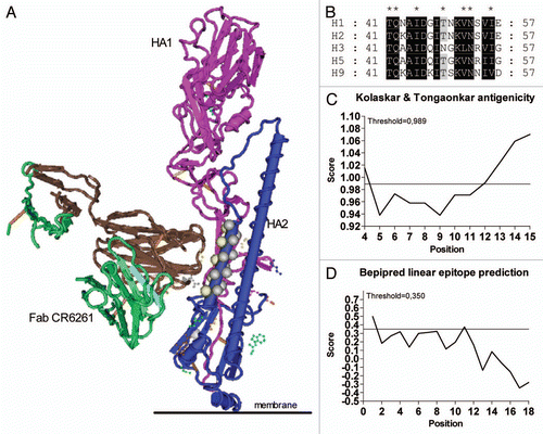
Figure 2 Immunization with HA2-KLH elicits potent anti-peptide immune response in bFcRn tg mice. Tg and wt mice were immunized with HA2 peptide conjugated to KLH in CFA and challenged in IFA on 21th and 42th day without adjuvant. Sera were analyzed for HA2 and KLH-specific IgG and IgM. (A) HA2-specific IgG titers showed a substantial increase in tg mice compared to the negligible IgG titers of wt mice even before the booster immunization. (B) HA2-specific IgM titers of tg mice were elevated during the secondary immune response compared to wt mice. Each circle represents an individual mouse. Lines represent the mean ± SEM. (*p < 0.05; **p < 0.01; ***p < 0.001). All the experiments were repeated twice with similar results.
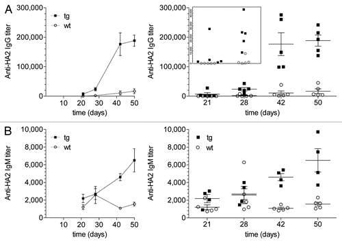
Figure 3 Both tg and wt mice showed strong immune response against the highly immunogenic KLH with three-fold difference in IgG titers in favor of the tg mice (A). We also detected increased KLH-specific IgM titers during the secondary immune response of tg mice. Each circle represents an individual mouse. Lines represent the mean ± SEM. All the experiments were repeated twice with similar results.
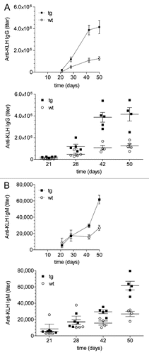
Figure 4 Immunization with HA2-KLH conjugate resulted in a difference between the spleen size of the animals with 2–3-fold increase in tg mice compared to wt mice (A) and also higher number of spleen cell number was observed in tg mice. Absolute number of B cells (C) and T cells (D) were significantly higher in the spleen of tg animals as measured by FACS analysis. Values shown are the mean ± SEM.
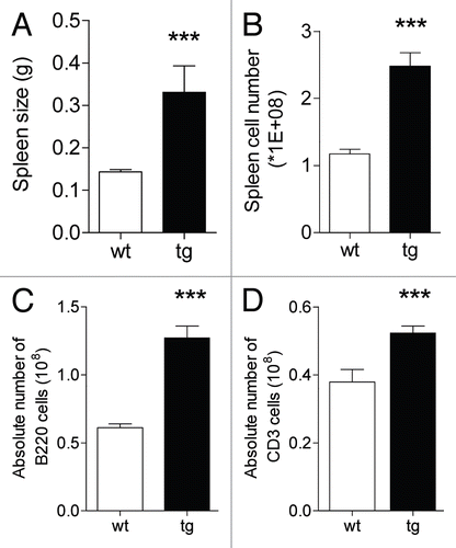
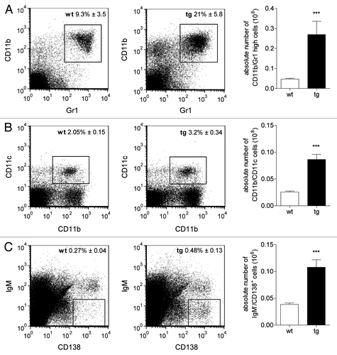
Figure 5 Differences were observed between the spleen cell populations of tg and wt mice after immunization as revealed by FACS analysis. Granulocytes, dendritic cells and IgM−/CD138+ cells (A–C, respectively) were present in significantly higher numbers in the spleen of tg mice. Absolute numbers of these cell types calculated based on the total spleen cell number showed multiple fold increase in tg animals. Values shown are the mean ± SEM.