Abstract
Antibody-drug conjugates (ADCs), formed through the chemical linkage of a potent small molecule cytotoxin (drug) to a monoclonal antibody, have more complex and heterogeneous structures than the corresponding antibodies. This review describes the analytical methods that have been used in their physicochemical characterization. The selection of the most appropriate methods for a specific ADC is heavily dependent on the properties of the linker, the drug, and the choice of attachment sites (lysines, inter-chain cysteines, Fc glycans). Improvements in analytical techniques such as protein mass spectrometry and capillary electrophoresis have significantly increased the quality of information that can be obtained for use in product and process characterization, and for routine lot release and stability testing.
Acknowledgements
The authors would like to thank Godfrey Amphlett, ImmunoGen, Inc., Michael Sun, Seattle Genetics, Inc. and Matt Kalo, Genentech, Inc. for providing original versions of figures.
Figures and Tables
Figure 1 Chemical structures of mAb-drug conjugates.Citation6 The linkers used for each drug are indicated in parentheses, and the labile bonds leading to drug release are shaded. For the examples shown, hydrazones release drug under acidic conditions within the lysozomes of target cells, disulfides undergo intracellular reduction, and the peptides are enzymatically hydrolyzed by lysozomal proteases. Adapted by permission from Macmillan Publishers Ltd. Wu AM, Senter PD. Arming antibodies: prospects and challenges for immunoconjugates.Citation6
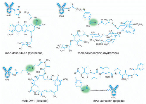
Figure 2 Hydrophobic interaction chromatography (HIC) analysis of a mAb-vc-MMAE on a TOSOH Biosciences Butyl-NPR column yields five predominant peaks that correspond to mAb containing zero, two, four, six and eight drugs. Inset: an overlay of the UV spectra of the starting mAb and the HIC peaks, normalized to the 280 nm absorbance, showing the increase in the 248 nm absorbance as the level of conjugated drug-linker increases. Method is similar to that described in Hamblett, et al.Citation23
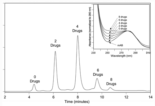
Figure 3 SEC/ESI-MS analysis of deglycosylated huC242-DM4 showing the deconvoluted mass spectrum. The label on each peak (e.g., D2) refers to the number of bound drug-linkers. Spectrum is obtained on a Waters LCT time of flight instrument as described by Lazar et al.Citation33 Inset: The total ion current in the SEC elution region of the protein. Adapted by permission from John Wiley & Sons: Lazar AC, Wang L, Blattler WA, Amphlett G, Lambert JM, Zhang W. Analysis of the composition of immunoconjugates using size-exclusion chromatography coupled to mass spectrometry.Citation33
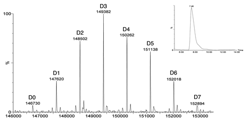
Figure 4 (A–D) Reversed-phase HPLC analysis of DTT-reduced conjugates produced using different reduction/reoxidation protocols. (E–H) Analysis of the same conjugate samples in (A–D), under non-reducing conditions, using the Agilent Bioanalyzer™, a silicon chip based system for capillary electrophoresis in the presence of SDS (CE-SDS).Citation12 Adapted with permission from Sun MM, Beam KS, Cerveny CG, Hamblett KJ, Blackmore RS, Torgov MY, et al. Reduction-alkylation strategies for the modification of specific monoclonal antibody disulfides.Citation12
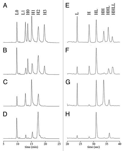
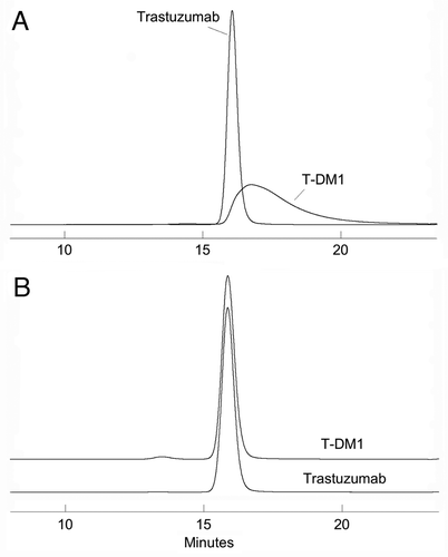
Figure 5 SEC analysis on a TSK 3000SWXL column run at 0.5 mL/min and monitored by 280 nm absorbance. (A) Mobile phase is 0.2 M KPi and 0.25 M KCl, pH 6.95. (B) 85% KPi/KCl mobile phase; 15% 2-propanol.