
Abstract
Subcutaneous (SC) delivery is a common route of administration for therapeutic monoclonal antibodies (mAbs) with pharmacokinetic (PK)/pharmacodynamic (PD) properties requiring long-term or frequent drug administration. An ideal in vivo preclinical model for predicting human PK following SC administration may be one in which the skin and overall physiological characteristics are similar to that of humans. In this study, the PK properties of a series of therapeutic mAbs following intravenous (IV) and SC administration in Göttingen minipigs were compared with data obtained previously from humans. The present studies demonstrated: (1) minipig is predictive of human linear clearance; (2) the SC bioavailabilities in minipigs are weakly correlated with those in human; (3) minipig mAb SC absorption rates are generally higher than those in human and (4) the SC bioavailability appears to correlate with systemic clearance in minipigs. Given the important role of the neonatal Fc-receptor (FcRn) in the PK of mAbs, the in vitro binding affinities of these IgGs against porcine, human and cynomolgus monkey FcRn were tested. The result showed comparable FcRn binding affinities across species. Further, mAbs with higher isoelectric point tended to have faster systemic clearance and lower SC bioavailability in both minipig and human. Taken together, these data lend increased support for the use of the minipig as an alternative predictive model for human IV and SC PK of mAbs.
Disclosure of Potential Conflicts of Interest
All authors are employees of Genentech, Inc., a member of the Roche Group or F. Hoffmann-La Roche and are Roche stockholders.
Acknowledgments
This study was supported financially by Genentech, Inc., a member of the Roche Group and F. Hoffmann-La Roche. We would like to thank Wayne Kung, Carolina Chou and Joseph Meyer for coordinating the minipig PK studies, Isabelle Bauer Dauphin, Ulla Grauschopf, Michael Adler, Sreedhara Alavattam, Jamie Moore, Mechelle Carnine, Osi Esue and Srikanth Chary for providing the dosing formulations, Sirj Goswami for carrying out part of the pharmacokinetic studies, David Michels for conducting the pI measurements, Zhenling Yao, Victor Yip and Crystal Zhang for their helpful discussions. We also would like to express appreciation to the key personnel involved in conducting the studies across the four contract research organizations (Pipeline Biotech, Trige, Denmark; Charles Rivers Labs, Ohio, USA; Covance Laboratories, Harrogate, UK; and LAB Research, Lille Skensved, Denmark).
Figures and Tables
Figure 1 Dose-normalized plasma or serum concentration-time curves following intravenous (closed symbols) or subcutaneous (open symbols) administration to Göttingen minipigs of (A) mAb1, (B) mAb2, (C) mAb3, (D) mAb4, (E) mAb5, (F) mAb6, (G) mAb7, (H) adalimumab and (I) mAb8 (intravenous dosing only) (mean ± SD).
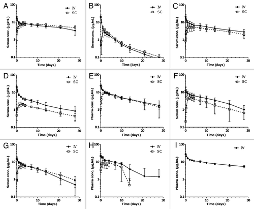
Figure 2 Correlation between minipig and human (A) clearance and (B) subcutaneous bioavailability following intravenous and subcutaneous administration of various monoclonal antibodies. Circles represent the reported mean parameter values while error bars represent the standard errors of the estimate; solid lines represent the linear regression lines (with r2 values displayed), and dashed lines represent the hypothetical line where the minipig and human values are identical. Note: in cases when more than one bioavailability value was reported, the average of all the reported values was used for the correlation analysis. For (A), mAb2 is excluded.
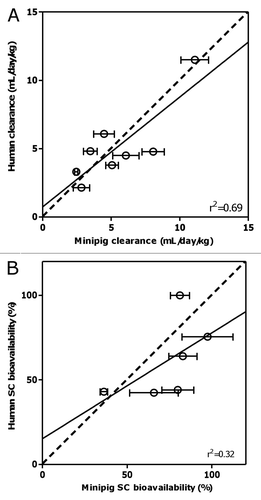
Figure 3 Correlation between clearance and subcutaneous bioavailability of the various mAbs in (A) minipig and (B) human. Black dots represent the reported mean parameter values while error bars represent the standard errors of estimate. For (A), mAb2 is excluded; solid line represents the linear regression line (with r2 value displayed).
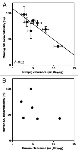
Figure 4 Correlation between pI and clearance in minipig (A) and human (B), and SC bioavailability in minipig (C) and human (D) of various monoclonal antibodies. Solid dots represent the reported mean parameter values while vertical error bars represent the standard errors of estimate. For (A), mAb2 is excluded. Grey dots represent mAb5 (outlier) whereas black dots represent all other mAbs.
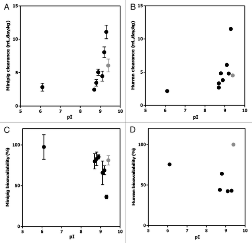
Figure 5 (A) Percent radioactivity remaining in the cell pellets following a 1 h incubation at 37°C of radioiodinated antibodies in minipig whole blood. (B) Size-exclusion HPLC radiochromatograms of [125I]-antibodies in plasma isolated from minipig whole blood following 1 h incubation.
![Figure 5 (A) Percent radioactivity remaining in the cell pellets following a 1 h incubation at 37°C of radioiodinated antibodies in minipig whole blood. (B) Size-exclusion HPLC radiochromatograms of [125I]-antibodies in plasma isolated from minipig whole blood following 1 h incubation.](/cms/asset/3cdcff8b-daae-4d18-8a9c-41c522c18c53/kmab_a_10919387_f0005.gif)
Table 1 Minipig studies: Pharmacokinetic parameters after intravenous and subcutaneous dosing for different monoclonal antibodies (mAbs)
Table 2 Allometric scaling of clearance for various monoclonal antibodies from minipigs to humans
Table 3 Human rate of absorption and bioavailability after subcutaneous dosing for different monoclonal antibodies
Table 4 Isoelectric points (pI) and in vitro FcRn binding affinities (KD) at pH 6 (mean and standard error)
Table 5 Experimental conditions of in vivo pharmacokinetic studies in Göttingen minipigs