
Abstract
Isolation of adipose-derived stem cells (ASCs) typically involves 8+ hours of intense effort, requiring specialized equipment and reagents. Here, we present an improved technique for isolating viable populations of mesenchymal stem cells from lipoaspirate saline fractions within 30 minutes. Importantly, the cells exhibit remarkable similarities to those obtained using the traditional isolation protocols, in terms of their multipotent differentiation potential and immunophenotype. Reducing the acquisition time of ASCs is critical for advancing regenerative medicine therapeutics, and our approach provides rapid and simple techniques for enhanced isolation and expansion of patient-derived mesenchymal stem cells.
Acknowledgements
We would like to thank Dr. Matthew Beckman for providing bone marrow mesenchymal stem cells. We are also grateful to Dr. Stephen Chen and his surgical team for facilitating accrual of lipoaspirates and surgical coordination. Flow Cytometry was supported in part by the Massey Cancer Center core grant, NIH Grant P30 CA16059. Microscopy was performed at the VCU—Dept. of Neurobiology & Anatomy Microscopy Facility, supported, in part, with funding from NIH-NINDS Center core grant (5P30NS047463).
Figures and Tables
Figure 1 Adipose-derived stem cell isolation techniques flow chart. The wide variance in the time, materials, and effort for obtaining ASCs via the Standard Isolation and our Rapid Isolation techniques is shown. A highly viable population of around 250,000 ASCs can be derived from 250 ml of blood/saline fraction of the liposuction waste in as little as 30 minutes, as compared to 8–10 hours to obtain ASCs using the traditional isolation method. SVF, stromal vascular fraction; ABAM, antibiotic/antimycotic; FBS, fetal bovine serum.
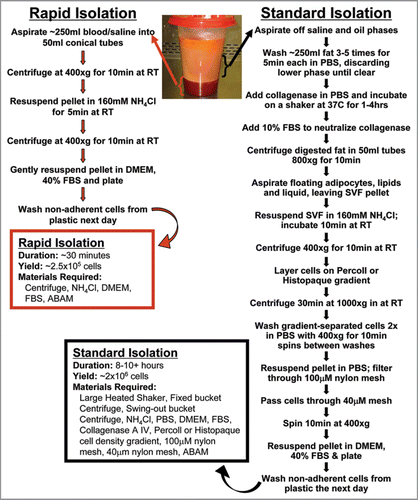
Figure 2 Characterization of ASC differentiation. The differentiation potential of ASCs isolated in the streamlined, rapid protocol were compared to ASCs isolated using the standard protocol (traditional ASCs). Cells were induced to differentiate into adipocytes ((A) for rapid ASCs and (G) for traditional ASCs; (D) rapid ASCs grown for 2 weeks without adipogenic media as control; all stained with oil red O and hematoxlin), osteocytes ((B) for rapid ASCs and (H) for traditional ASCs; (E) rapid ASCs grown 2 weeks without osteocyte induction media as control; all stained with Alizarin red S and hematoxylin), and chondrocytes [(C) for rapid ASCs and (I) for traditional ASCs, both in a micromass (solid micromass pellet, insert, (C)]; (F) unpelleted rapid ASCs grown 4 weeks in induction media as control; all samples stained for Safranin O).
![Figure 2 Characterization of ASC differentiation. The differentiation potential of ASCs isolated in the streamlined, rapid protocol were compared to ASCs isolated using the standard protocol (traditional ASCs). Cells were induced to differentiate into adipocytes ((A) for rapid ASCs and (G) for traditional ASCs; (D) rapid ASCs grown for 2 weeks without adipogenic media as control; all stained with oil red O and hematoxlin), osteocytes ((B) for rapid ASCs and (H) for traditional ASCs; (E) rapid ASCs grown 2 weeks without osteocyte induction media as control; all stained with Alizarin red S and hematoxylin), and chondrocytes [(C) for rapid ASCs and (I) for traditional ASCs, both in a micromass (solid micromass pellet, insert, (C)]; (F) unpelleted rapid ASCs grown 4 weeks in induction media as control; all samples stained for Safranin O).](/cms/asset/9ee2896e-21bb-4eb5-a725-95d2e6159d54/kogg_a_10910019_f0002.gif)
Table 1 Immunophenotype of ASCs isolated using the rapid and standard protocols
Table 2 EGF supplemented ASC media enhances growth