
Abstract
A simple diagnostic test is described for the detection of TSE in bovine, ovine and human brain and lymphoid tissue that obviates the use of proteinase K as a discriminating reagent. The immunoassay utilises high affinity anti-peptide antibodies that appear blind to the normal isoform of prion protein (PrPC). These reagents have been produced with novel N-terminal chimeric peptides and we hypothesise that the retention and stability of the extreme N-terminus of PrP in the disease-associated aggregate makes it an operationally specific marker for TSE. Accordingly, the assay involves homogenisation of the tissue directly in 8M guanidine hydrochloride, a simple one-step capture of PrPSc followed by detection with a europium-labelled anti-PrPC antibody. This rapid assay clearly differentiates between levels of disease-associated PrP extracted from brain and lymphoid tissues taken from confirmed TSE positive and negative cattle and sheep. The assay can also be used to detect PrPSc in cases of vCJD.
Acknowledgements
The authors would like to thank VLA for their support and encouragement, particularly in the provision of confirmed positive and negative tissues. Without their support, none of this work could have been accomplished. In particular, we would like to acknowledge our colleagues at the VLA in Newcastle for the opportunity to perform aspects of the work using their facilities. In addition, we are grateful to PerkinElmer Life Sciences for their support and for the provision of equipment and reagents. We thank Drs. Raymond Bujdoso and Alana Thackray for constructive discussions and TJ McKinley, Department of Veterinary Medicine, Madingley Road, Cambridge CB3 0ES, UK for expert advice regarding the statistical analysis of the data. We are grateful to the TSE Joint Funders for financial support (BBSRC Grants 8/BSD17730 and BB/D004500).
Conflict of Interest/Financial Disclosure Statement
The work described in this paper is subject to the UK Patent Application (PCT/GB2006/003494). This patent application, however, has not influenced our scientific judgment.
Figures and Tables
Figure 1 N-terminal bovine (A) and human (B) chimeric peptide used as immunogens for the production of YWH antibodies.
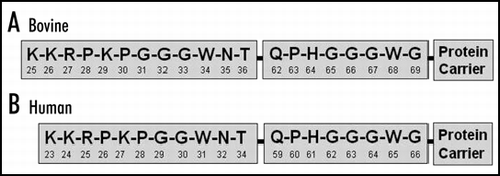
Figure 2 Cross reactions of YWH1 and YWH2 using various peptides.
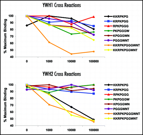
Figure 3 Measurement of disease-associated PrP in histology-confirmed scrapie positive and negative rostral medulla using Western blot and DELFIA®.
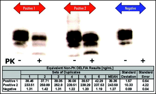
Figure 4 Measurement of disease-associated PrP in vCJD-spiked human tonsil homogenate.
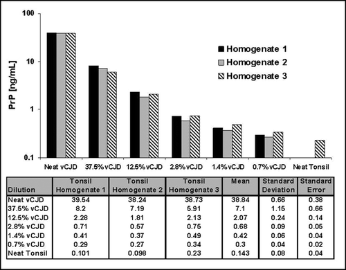
Table 1 Measurement of disease-associated PrP in histology-confirmed BSE positive and negative bovine caudal medulla.
Table 2 Measurement of disease-associated PrP in histology-confirmed Scrapie positive and negative ovine caudal medulla (A), rostral medulla (B), palatine tonsil (C) and retropharyngeal lymph node (D)