Abstract
The absence of specific immune response is a hallmark of prion diseases. However, in vitro and in vivo experiments have provided evidence that an anti-PrP humoral response could have beneficial effects. Prophylactic passive immunization performed at the time of infection delayed or prevented disease. Nonetheless, the potential therapeutic effect of PrP antibodies administered shortly before the clinical signs has never been tested in vivo. Moreover, a recent study showed the potential toxicity of PrP antibodies administered intracerebrally. We aimed at evaluating the effect of a prolonged intracerebral anti-PrP antibody administration at the time of neuroinvasion in BSE infected Tg20 mice. Unexpectedly, despite a good penetration of the antibodies in the brain parenchyma, the treatment was not protective against the development of BSE. Instead, it led to an extensive neuronal loss, strong astrogliosis and microglial activation. Since this effect was observed after injection of anti-PrP antibodies as whole IgGs, F(ab')2 or Fab fragments, the toxicity was directly related to the ability of the antibodies to recognize native PrP and to the intracerebral concentration achieved, and not to the Fc portion or the divalence of the antibodies. This experiment shows that a prolonged treatment with anti-PrP antibodies by the intracerebral route can induce severe side-effects and calls for caution with regard to the use of similar approaches for late therapeutic interventions in humans.
Acknowledgments
This work was financially supported by the Commissariat à L'Energie Atomique (CEA, Fontenay-aux-Roses, France), the Agence Française de Sécurité Sanitaire des Aliments (AFSSA, Lyon, France), the Scripps Research Institute (Jupiter, Florida, USA) and by the European Community (grant ≠ QLK3-CT-2001-00283). We are indebted to Drs. G. Kneale and J. McGeehan (University of Portsmouth, UK) for having kindly provided recombinant g5p protein. We are grateful to J. Comte, B. Dufresnois, J. Desmercière and M.C. Mondon for helpful technical assistance. We gratefully acknowledge the critical reading of the manuscript by C. Weissmann.
Figures and Tables
Figure 1 Distribution of the antibodies in the brain. The antibodies were detected in the brain of the infected animals after a continuous 15 days treatment during the neuroinvasion (85–100 dpi). Red-brown labeling of the antibodies in the encephalon (first row, bar = 2.5 mm) highlights the greater diffusion rate of the F(ab′)2 fragments (columns 4 and 5) when compared to the corresponding IgGs (columns 2 and 3). Higher magnification microphotographs of the thalamus (second row, bar = 50 µm) reveal the distribution of the antibodies in the neuropile and in cell bodies. The mice treated with 4H11 F(ab′)2 fragments displayed a strong perturbation of the neuropile associated with shrunk cells and pycnotic nuclei (column 4).
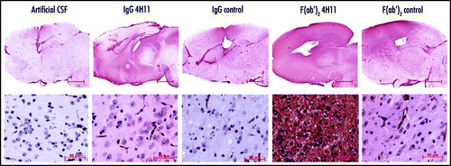
Figure 2 Survival of 6PB1 infected mice. Grey circles: mice infused with the artificial CSF. Black triangle: mice treated with the PrP and control IgGs (left panel). Black squares: mice treated with PrP and control F(ab′)2 fragments (right panel).
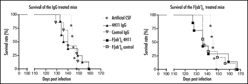
Figure 3 PrPSc accumulation in the brain of treated animals. (A) PrPSc was enriched from brain homogenates by precipitation with g5p and digested or not with PK (-/+ lanes). PrPSc was evaluated at the end of the treatement (lanes 1–10) or at the terminal stage of disease (lanes 11 and 12). (B) PrPSc was purified from brains of animals at the end of treatments (left set of bars), or at terminal stage (right set of bars) by the purification technique described in the methods, and then detected by sandwich ELISA.
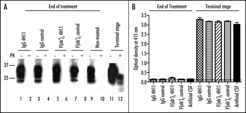
Figure 4 Effect of the antibody treatment on neurons and glial cells in infected animals at the end of the treatment. (A) Neuron specific NeuN labeling highlighting the neuronal loss in the regions CA1 and CA2 of the hippocampus of the animals treated with 4H11 F(ab′)2 fragments, while the animals from all the other groups did not show neuronal loss (row 1). TUNEL labeling showing apoptotic neurons in the same region of the hippocampus of the animals treated with 4H11 F(ab′)2 fragments (row 2). Bar = 500 µm. (B) Higher magnification and labeling for gliosis of the hippocaampus of the 4H11 F(ab′)2 treated mouse shown in (A) (lines show the corresponding panel A). Immunofluorescence analysis revealed the presence of GFAP positive reactive astrocytes (row 1, red labeling) and F4/80 positive microglial cells (row 2, red labeling) in the vicinity of the apoptotic neurons labelled by TUNEL (2nd and 4th columns, green) in the hippocampus of 4H11 F(ab′)2 fragments treated animals. Bar = 50 µm.
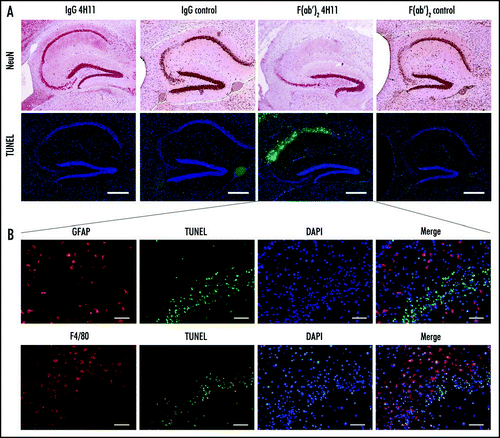
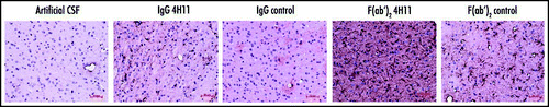
Figure 5 Effect of the antibody treatment on the astroglial reaction. Reactive astrocytes were detected at the end of the treatment by labeling for GFAP (brown). (Thalamus, Bar = 50 µm).