
Abstract
The structure and the dissociation reaction of oligomers PrPoligo from reduced human prion huPrPC (23-231) have been studied by 1H-NMR and tryptophan fluorescence spectroscopy at varying pressure, along with circular dichroism and atomic force microscopy. The 1H-NMR and fluorescence spectral feature of the oligomer is consistent with the notion that the N-terminal residues including all seven Trp residues, are free and mobile, while residues 105~210, comprising the AGAAAAGA motif and S1-Loop-HelixA-Loop-S2-Loop-HelixC, are engaged in intra- and/or inter-molecular interactions. By increasing pressure to 200 MPa, the oligomers tend to dissociate into monomers which may be identified with PrPC*, a rare metastable form of PrPC stabilized at high pressure (Kachel et al. BMC. Struct. Biol. 6, 16). The results strongly suggest that the oligomeric form PrPoligo is in dynamic equilibrium with the monomeric forms via PrPC*, namely huPrPC ⇄ huPrPC* ⇄ huPrPoligo.
Acknowledgements
This work was supported by the BSE Control Project from MAFF (the Ministry of Agriculture, Forestry and Fisheries of Japan) and by a Grant-in-Aid for Scientific Research No. 16370054 from MEXT (the Ministry of Education, Culture, Sports, Science and Technology of Japan), and carried out under the sponsorship of an Academic Frontier Program of MEXT. The German-Japanese cooperation was supported by the DFG.
Figures and Tables
Figure 1 Circular dichroism spectra of monomeric oxidized huPrPC(23–231) (blue) and oligomeric reduced huPrPC(23–231) (red), recorded at 298 K. Monomeric oxidized huPrPC(23–231) (0.12 mM) was prepared in a buffer solution containing 10 mM sodium acetate and 10 mM tris-acetate, pH 8.0 and oligomeric reduced huPrPC(23–231) (1.9 mM) was prepared in a buffer solution containing 10 mM sodium acetate, 10 mM tris-acetate, pH 4.0.
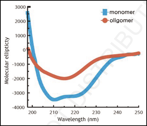
Figure 2 Atomic force microscopic image of oligomeric reduced huPrPC(23–231), recorded at room temperature.
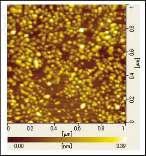
Figure 3 Trp fluorescence spectra of oligomeric reduced huPrPC(23–231) recorded at varying pressures and at 298 K.
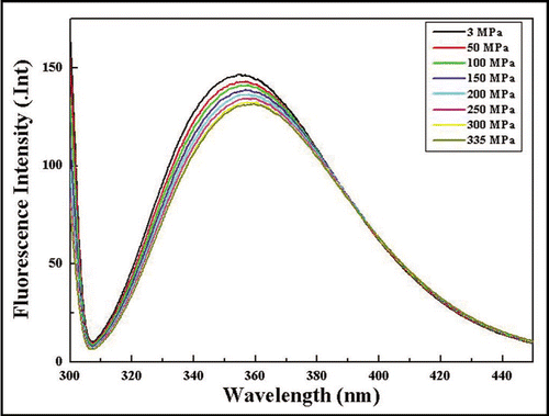
Figure 4 1H-NMR spectral changes of monomeric and oligomeric huPrPC(23–231) caused by pressure-jump up and down. (A) 1H NMR spectrum of (15N-enriched) monomeric huPrPC(23–231) at 293 K at 0.3 MPa. (B) The same as (A), but at 200 MPa. (C) 1H NMR spectrum of reduced oligomeric huPrPoligo(23–231) at 298 K at 0.3 MPa. (D) The same as (C), but 30 min after the pressure-jump from 0.3 MPa to 200 MPa. (E) The same as (C), but 46 h after the pressure-jump from 0.3 MPa to 200 MPa. (F) The same as (C), but 30 min after the pressure-jump back from 200 MPa to 0.3 MPa. (G) The same as (C), but 9.5 h after the pressure-jump back from 200 MPa to 0.3 MPa.
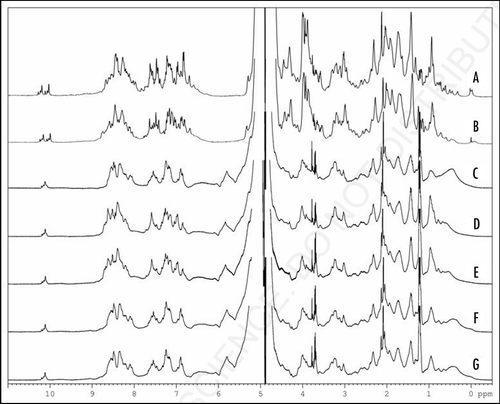
Figure 5 Oligomeric and monomeric of huPrPC 1H NMR recorded at varying pressures. (A) 1H NMR spectra of huPrPC(23–230) recorded at 293 K, pH 4.8 and at 0.1 MPa. Resonances of selected protons are labeled: (1) Leu130 Hδ2 (2) Leu125 Hδ2 (3) Ile139 Hδ1 (4) Ile182 Hδ1 (5) Ile182 Hγ2 (6) DSS (internal reference) (7) Leu130 Hδ1 (8) Ile139 Hγ2. (B) 1H NMR spectra of huPrPC(23–230) recorded at 293 K, pH 4.8 and at 200 MPa. [(A and B) data were reprinted from ref. Citation8]. (C) 1H NMR spectra of huPrPoligo(23–231) recorded at 298 K, pH 4.0 and at 0.3 MPa. (D) 1H NMR spectra of huPrPoligo(23–231) recorded at pressure jump up to 200 MPa after 46 h and at 298 K, pH 4.0. (E) 1H NMR spectra of huPrPoligo(23–231) recorded at pressure jump back to 0.3 MPa after 9.5 h and at 298 K, pH 4.0.
![Figure 5 Oligomeric and monomeric of huPrPC 1H NMR recorded at varying pressures. (A) 1H NMR spectra of huPrPC(23–230) recorded at 293 K, pH 4.8 and at 0.1 MPa. Resonances of selected protons are labeled: (1) Leu130 Hδ2 (2) Leu125 Hδ2 (3) Ile139 Hδ1 (4) Ile182 Hδ1 (5) Ile182 Hγ2 (6) DSS (internal reference) (7) Leu130 Hδ1 (8) Ile139 Hγ2. (B) 1H NMR spectra of huPrPC(23–230) recorded at 293 K, pH 4.8 and at 200 MPa. [(A and B) data were reprinted from ref. Citation8]. (C) 1H NMR spectra of huPrPoligo(23–231) recorded at 298 K, pH 4.0 and at 0.3 MPa. (D) 1H NMR spectra of huPrPoligo(23–231) recorded at pressure jump up to 200 MPa after 46 h and at 298 K, pH 4.0. (E) 1H NMR spectra of huPrPoligo(23–231) recorded at pressure jump back to 0.3 MPa after 9.5 h and at 298 K, pH 4.0.](/cms/asset/de1a58f6-03f7-4018-b5bf-7a6e6300b5b0/kprn_a_10907148_f0005.gif)