Abstract
Prion diseases are emerging infectious disorders that affect several mammalian species including humans. The transmissible agent is comprised of PrPSc, a misfolded isoform of the normal host-encoded prion protein PrPC. Immunodetection of PrPSc is often utilized for prion disease diagnosis and tracking spread of the infectious agent through the host. We have developed a rapid, high-throughput 96 well immunoassay, which is specific for the detection of PrPSc. This assay has PrPSc detection limits similar to Western blot and is advantageous because of its comparatively shorter running time, smaller start-up and operation costs, and large sample capacity.
Acknowledgements
This work was supported by the National Center for Research Resources (P20 RR0115635-6 and C06 RR17417-01), the National Institute for Neurological Disorders and Stroke (R01 NS052609) and the National Prion Research Program (NP020041).
Figures and Tables
Figure 1 96-well PrPSc immunoassay controls. (A) 96-well immunoassay and (B) quantification of brain homogenate from proteinase K digested uninfected (UN) or HY TME-agent infected (HY TME) hamsters with (+) and without (−) H2O2 in MeOH, primary antibody or secondary antibody to indicate the specificity of PrPSc immunodetection. Relative PrPSc intensity is based on well 16.
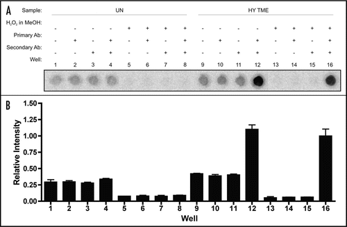
Figure 2 Limits of PrP detection using western blot analysis. Western blot analysis of two-fold serial dilutions of brain homogenates treated with (+) and without (−) proteinase K (PK) from uninfected hamsters (UN, A) or hamsters infected with the HY TME (HY) or DY TME (DY) agents (B and C, respectively).

Figure 3 Limits of PrP detection using 96-well immunoassay. 96 well immunoassay of two-fold serial dilutions of brain homogenate from uninfected (UN), HY TME agent (HY) or DY TME agent (DY) infected hamsters. Samples were either proteinase K (PK) digested (+) or were left untreated (−).
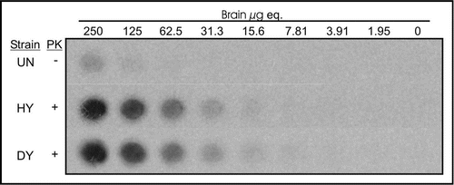
Figure 4 Comparison of PrPSc detection limits by western blot and 96-well immunoassay. Quantification of PrPSc abundance of two-fold serial dilutions of uninfected (UN), HY TME-infected (HY) or DY TME-infected (DY) proteinase K digested brain material by either western blot (open bars) or 96-well immunoassay (solid bars). Represented data is the average and standard error of the mean of three independent experiments.
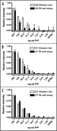
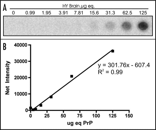
Figure 5 Linear range of PrPSc detection by 96-well immunoassay. (A) 96-well immunoassay and (B) quantification of PrPSc from 2 fold serial dilutions of HY TME-agent infected brain homogenate digested with proteinase K.